Copyright
©2009 The WJG Press and Baishideng.
World J Gastroenterol. Jul 14, 2009; 15(26): 3269-3275
Published online Jul 14, 2009. doi: 10.3748/wjg.15.3269
Published online Jul 14, 2009. doi: 10.3748/wjg.15.3269
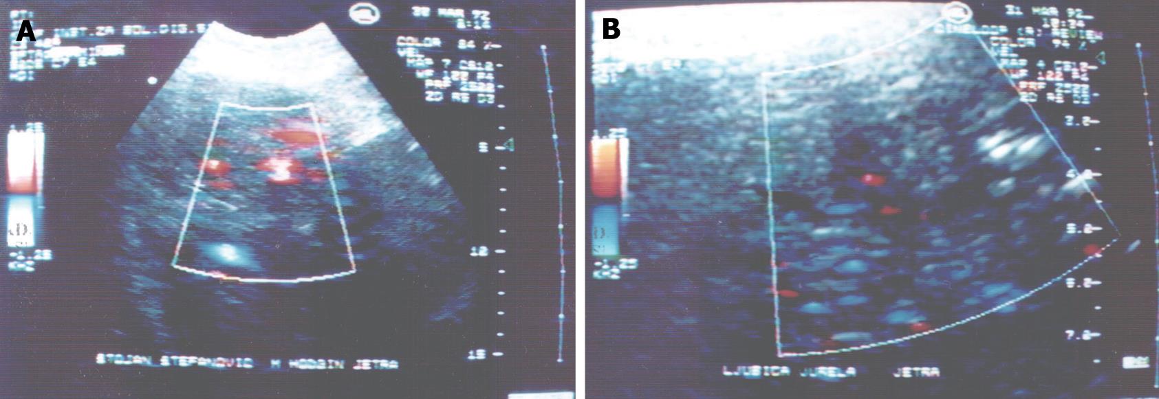
Figure 1 Color doppler ultrasonography.
A: Intense-central neovascularization in the focal liver lesion; B: Intense peripheral neovascularization in the focal liver lesion.
- Citation: Stojković MV, Artiko VM, Radoman IB, Knežević SJ, Lukić SM, Kerkez MD, Lekić NS, Antić AA, Žuvela MM, Ranković VI, Petrović MN, Šobić DP, Obradović VB. Color Doppler sonography and angioscintigraphy in hepatic Hodgkin’s lymphoma. World J Gastroenterol 2009; 15(26): 3269-3275
- URL: https://www.wjgnet.com/1007-9327/full/v15/i26/3269.htm
- DOI: https://dx.doi.org/10.3748/wjg.15.3269


