Copyright
©2009 The WJG Press and Baishideng.
World J Gastroenterol. May 21, 2009; 15(19): 2425-2427
Published online May 21, 2009. doi: 10.3748/wjg.15.2425
Published online May 21, 2009. doi: 10.3748/wjg.15.2425
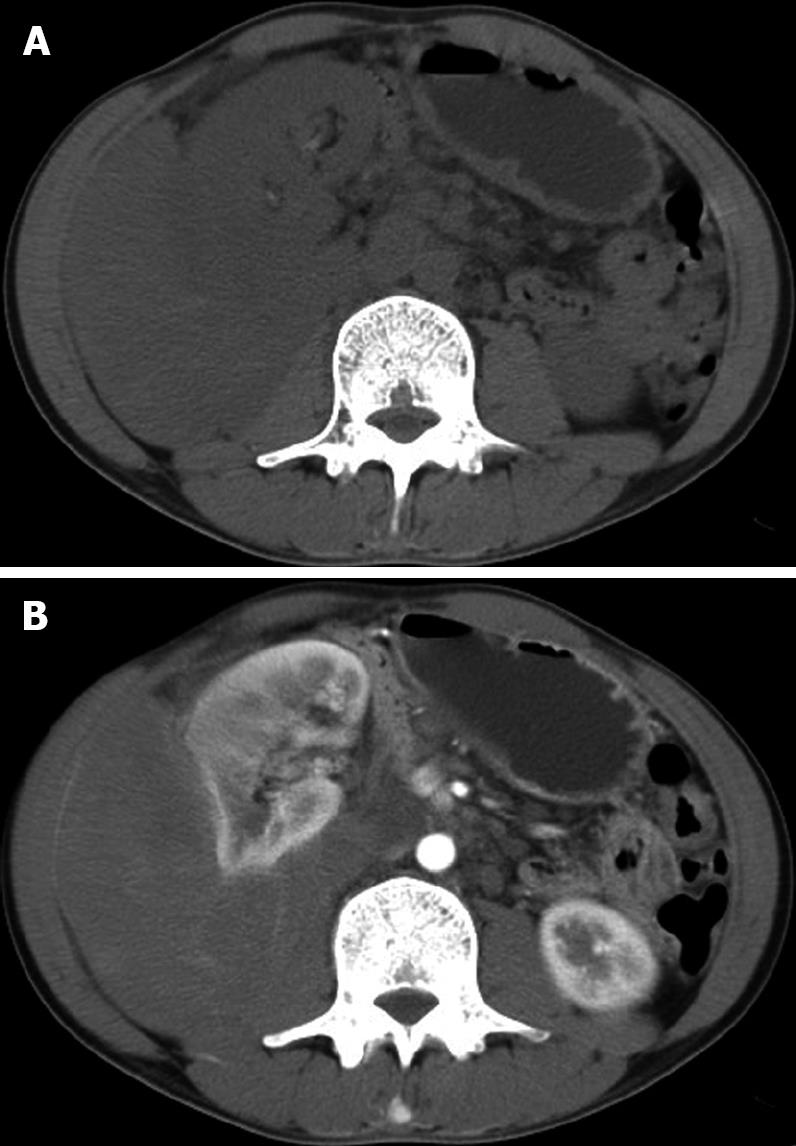
Figure 1 Abdominal computed tomography showing a large heterogeneous mass in the right retroperitoneal region, surrounding the posterior portion of the right kidney and compressing the right kidney (A), and slightly enhanced tumor tissue after injection of a contrast medium (B).
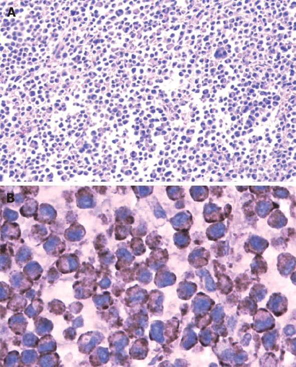
Figure 2 Tumor cells with homogenous amphophilic cytoplasm, whell-type and asymmetric nuclei, coarsely stippled chromatin, some acidophilic nucleoli, and occasional binucleate (A) (HE, × 100) and positive tumor cells for CD138 (B) (× 400) under microscope.
- Citation: Hong W, Yu XM, Jiang MQ, Chen B, Wang XB, Yang LT, Zhang YP. Solitary extramedullary plasmacytoma in retroperitoneum: A case report and review of the literature. World J Gastroenterol 2009; 15(19): 2425-2427
- URL: https://www.wjgnet.com/1007-9327/full/v15/i19/2425.htm
- DOI: https://dx.doi.org/10.3748/wjg.15.2425


