Copyright
©2009 The WJG Press and Baishideng.
World J Gastroenterol. May 21, 2009; 15(19): 2423-2424
Published online May 21, 2009. doi: 10.3748/wjg.15.2423
Published online May 21, 2009. doi: 10.3748/wjg.15.2423
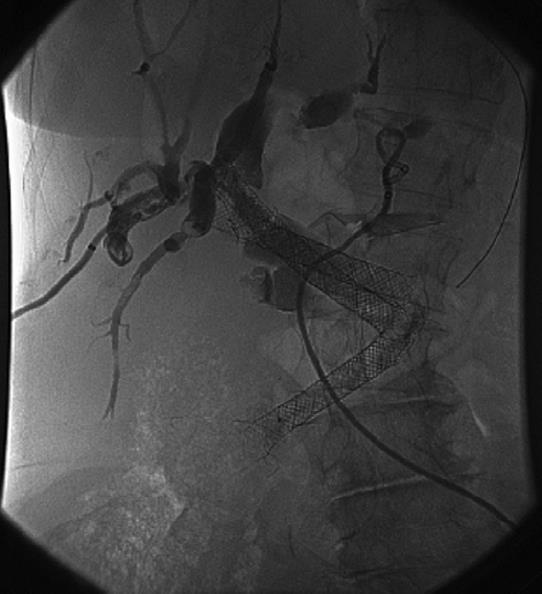
Figure 1 Radiography image.
At time of presentation, sludge and stones in the proximal bile ducts with five metal stents in the CBD (four proximally and one distally) and two externally draining 8 French percutaneous drains.
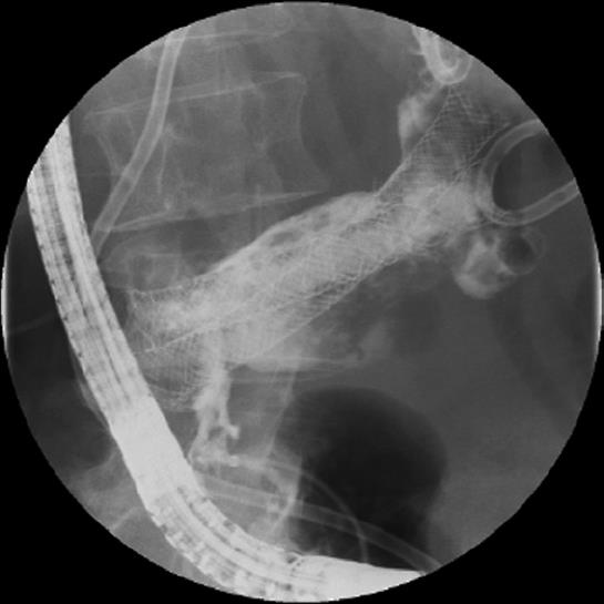
Figure 2 After endoscopic removal of the distal metal stent.
The distal CBD was visualized with 4 metal stents proximally in an abnormal position in relation to the CBD.
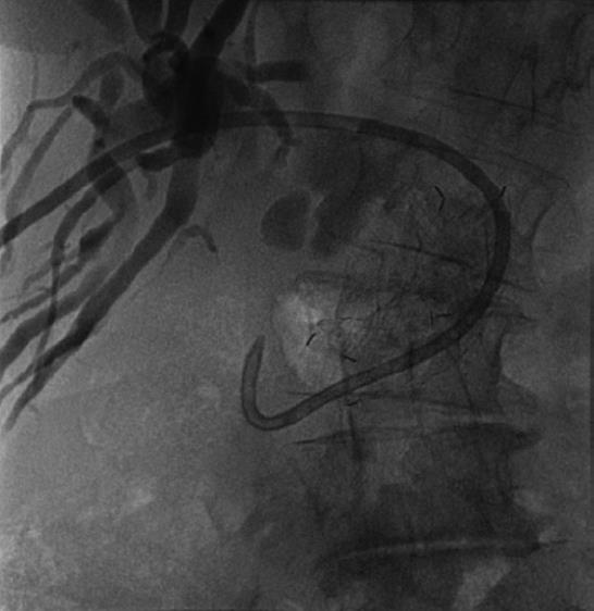
Figure 3 After placement of an enteral metal stent in the dilated CBD.
- Citation: Dek IM, van den Elzen BD, Fockens P, Rauws EA. Biliary drainage of the common bile duct with an enteral metal stent. World J Gastroenterol 2009; 15(19): 2423-2424
- URL: https://www.wjgnet.com/1007-9327/full/v15/i19/2423.htm
- DOI: https://dx.doi.org/10.3748/wjg.15.2423


