Copyright
©2009 The WJG Press and Baishideng.
World J Gastroenterol. May 14, 2009; 15(18): 2258-2264
Published online May 14, 2009. doi: 10.3748/wjg.15.2258
Published online May 14, 2009. doi: 10.3748/wjg.15.2258
Figure 1 Representative photograph of the TMA slides with immunohistochemical staining.
A: CD133; B: CD24; C: CD44.
Figure 2 Representative photographs of CD marker expression in colorectal adenocarcinoma.
Positive staining (A) and negative staining (B) for CD133. Positive staining (C) and negative staining (D) for CD24. Positive staining (E) and negative staining (F) for CD44.
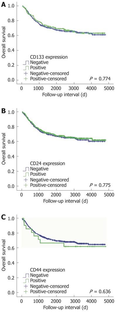
Figure 3 Cumulative survivals according to CD133 (P = 0.
774) (A), CD24 (P = 0.775) (B) and CD44 (P = 0.636) (C) expression in colorectal cancer patients (Kaplan-Meier method).
- Citation: Choi D, Lee HW, Hur KY, Kim JJ, Park GS, Jang SH, Song YS, Jang KS, Paik SS. Cancer stem cell markers CD133 and CD24 correlate with invasiveness and differentiation in colorectal adenocarcinoma. World J Gastroenterol 2009; 15(18): 2258-2264
- URL: https://www.wjgnet.com/1007-9327/full/v15/i18/2258.htm
- DOI: https://dx.doi.org/10.3748/wjg.15.2258


