Copyright
©2009 The WJG Press and Baishideng.
World J Gastroenterol. May 7, 2009; 15(17): 2166-2169
Published online May 7, 2009. doi: 10.3748/wjg.15.2166
Published online May 7, 2009. doi: 10.3748/wjg.15.2166
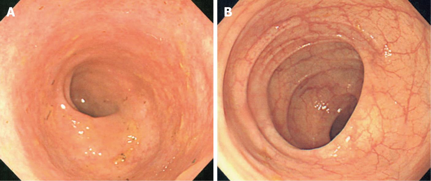
Figure 1 Colonoscopy on April 16 (A) and May 17 (B), 2007 showed diffuse cloudiness of mucosa in the colon and clear normal vascular patterns, respectively.
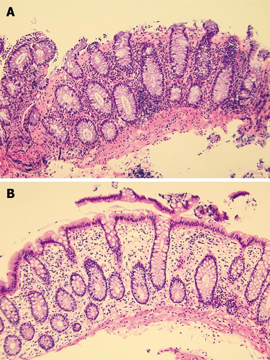
Figure 2 Biopsy specimens taken on April 16 (A) and May 17 (B), 2007 (hematoxylin and eosin staining, × 100).
The former showed erosion, moderate infiltration of inflammatory cells in the lamina propria, and subepithelial collagenous thickening. The latter showed disappearance of these abnormalities.
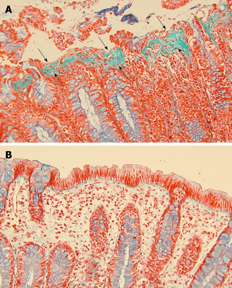
Figure 3 Biopsy specimens taken on April 16 (A) and May 17 (B), 2007 (Masson’s trichrome staining, × 200).
Subepithelial collagenous thickening (A, arrows) disappeared on May 17 (B).
- Citation: Chiba M, Sugawara T, Tozawa H, Tsuda H, Abe T, Tokairin T, Ono I, Ushiyama E. Lansoprazole-associated collagenous colitis: Diffuse mucosal cloudiness mimicking ulcerative colitis. World J Gastroenterol 2009; 15(17): 2166-2169
- URL: https://www.wjgnet.com/1007-9327/full/v15/i17/2166.htm
- DOI: https://dx.doi.org/10.3748/wjg.15.2166


