Copyright
©2009 The WJG Press and Baishideng.
World J Gastroenterol. Apr 14, 2009; 15(14): 1769-1770
Published online Apr 14, 2009. doi: 10.3748/wjg.15.1769
Published online Apr 14, 2009. doi: 10.3748/wjg.15.1769
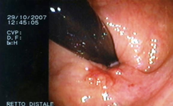
Figure 1 Endoscopic view of a polypoid, submucous, ulcerated lesion in its vertex.
Figure 2 Endoanal ultrasound scan showing a mass located in the anterior wall of the rectum.
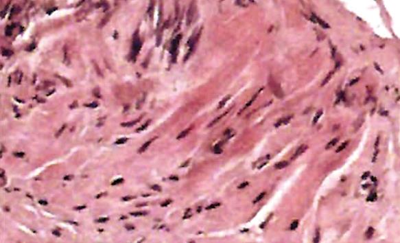
Figure 3 Microscopic findings showing a proliferation of fusiform, elongated spindle cells arranged in fascicles.
- Citation: Palma GDD, Rega M, Masone S, Siciliano S, Persico M, Salvatori F, Maione F, Esposito D, Bellino A, Persico G. Lower gastrointestinal bleeding secondary to a rectal leiomyoma. World J Gastroenterol 2009; 15(14): 1769-1770
- URL: https://www.wjgnet.com/1007-9327/full/v15/i14/1769.htm
- DOI: https://dx.doi.org/10.3748/wjg.15.1769


