Copyright
©2009 The WJG Press and Baishideng.
World J Gastroenterol. Apr 14, 2009; 15(14): 1665-1672
Published online Apr 14, 2009. doi: 10.3748/wjg.15.1665
Published online Apr 14, 2009. doi: 10.3748/wjg.15.1665
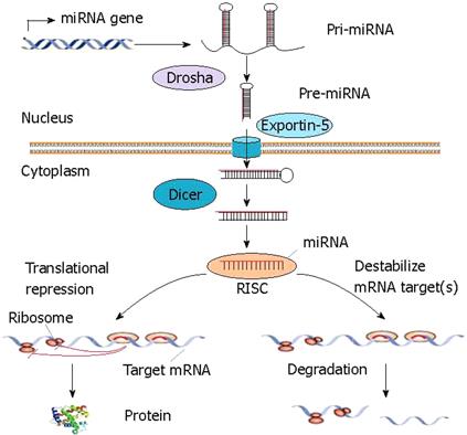
Figure 1 miRNA biogenesis and function in animal cells.
Animal genomes have specific genes that encode miRNAs. Primary miRNA transcripts (pri-miRNAs) are processed into precursor miRNA (pre-miRNAs) stem-loops of approximately 60 nucleotides in length by the nuclear RNase III enzyme Drosha. These pre-miRNAs are transported to the cytoplasm via exportin-5 and are further processed by the ribonuclease Dicer. Mature miRNAs are then incorporated in the RNA-induced silencing complex (RISC) and interfere with the regulation of mRNA translation by targeting mRNAs resulting in mRNA degradation or translational repression.
Figure 2 Prediction of targets by miRNAs in the liver.
The miRNAs are ordered by RR values (upper-axis). The higher values reflect lower expression of predicted target genes and are, therefore, indicative of miRNA activity. The numbers of genes predicted to be targeted by each miRNA (lower-axis) are indicated by the grey line. This figure is reprinted from Arora and Simpson[28] with the permission of the authors and the publisher.
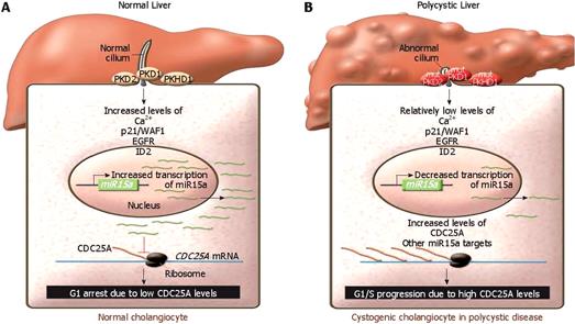
Figure 3 Model of miR15a function in hepatic cystogenesis.
A: In normal cholangiocytes, ciliary signaling activates intracellular signaling pathways resulting in miR15a expression and CDC25A repression. Decreased levels of CDC25A then lead to G1 arrest, preventing cyst formation; B: In cholangiocytes of polycystic liver, miR15a level is reduced as a result of mutation of molecules important to the ciliary signaling. Reduction of miR15a results in elevated Cdc25A level, increased cell proliferation, and cyst growth. This figure is reprinted from Chu and Friedman[49] with the permission of the authors and the publisher.
- Citation: Chen XM. MicroRNA signatures in liver diseases. World J Gastroenterol 2009; 15(14): 1665-1672
- URL: https://www.wjgnet.com/1007-9327/full/v15/i14/1665.htm
- DOI: https://dx.doi.org/10.3748/wjg.15.1665


