Copyright
©2008 The WJG Press and Baishideng.
World J Gastroenterol. Feb 28, 2008; 14(8): 1167-1174
Published online Feb 28, 2008. doi: 10.3748/wjg.14.1167
Published online Feb 28, 2008. doi: 10.3748/wjg.14.1167
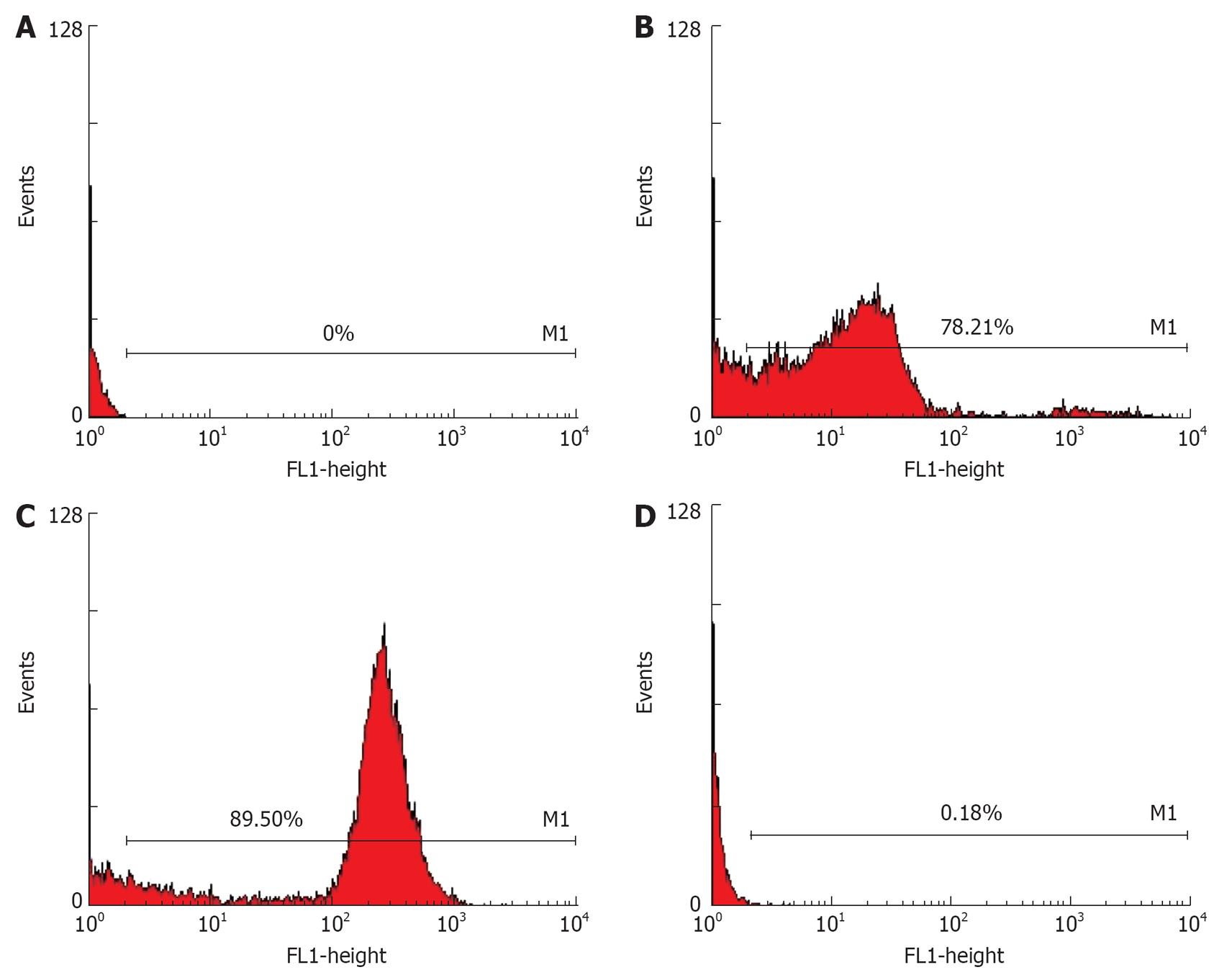
Figure 1 Expression of folate receptor on EC109 and DCs.
PBL (A) was set up for negative cell and HNE1 for masculine cell. Analyses by flow cytometry, HNE1 (B) with expression of FR was 78.21%, EC109 (C) was 89.50%, and DCs (D) was 0.18%.
Figure 2 Expression of FR (A), CD80 (B) and EC109-DC (C).
Figure 3 No formation of tumor in the liver (A), in the kidney 60 d after injection of EC109-DC into the vena caudalis (B) (HE, 20 × 10) and in tumor tissue (C) of SCID mice 28 d after injection of EC109 into the abdominal cavity (HE, 10 × 10).
Figure 4 Incubation time (A), weight (B) and diameter (C) of tumors after attacked by EC109.
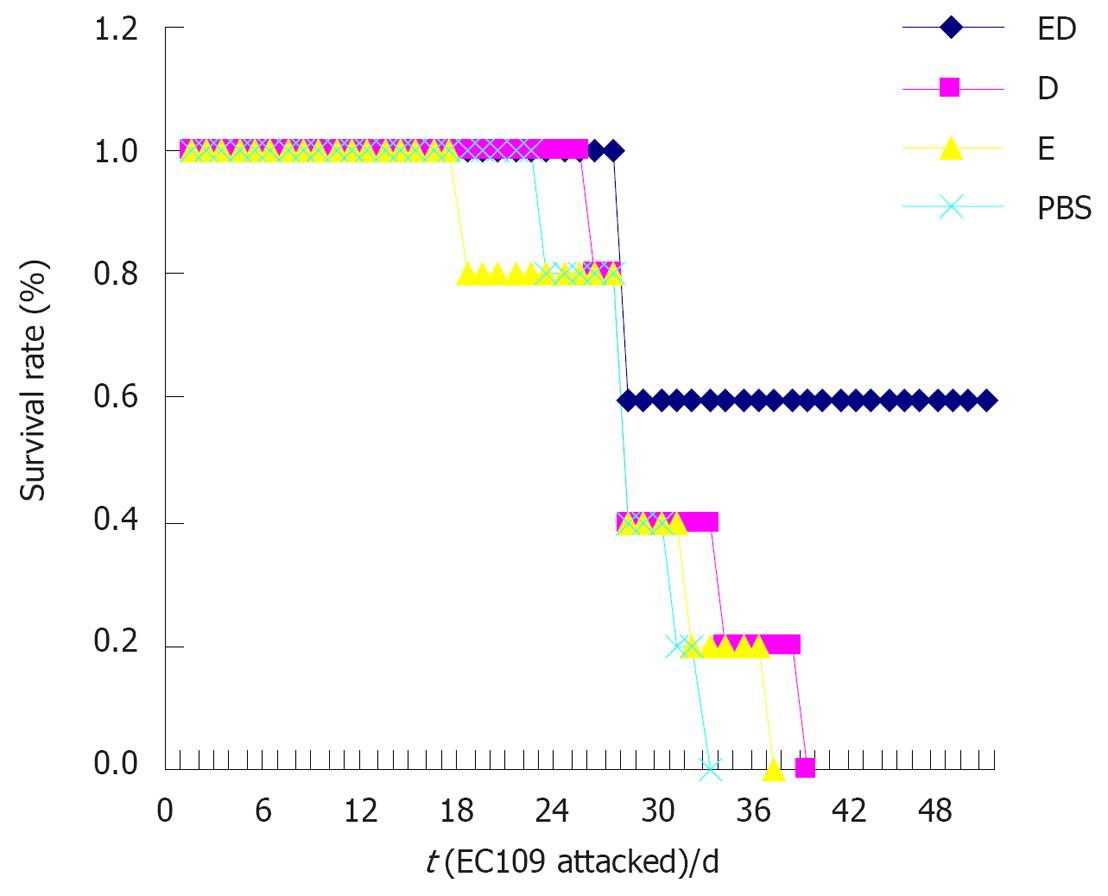
Figure 5 Death and life span in immune, ED and PBS groups.
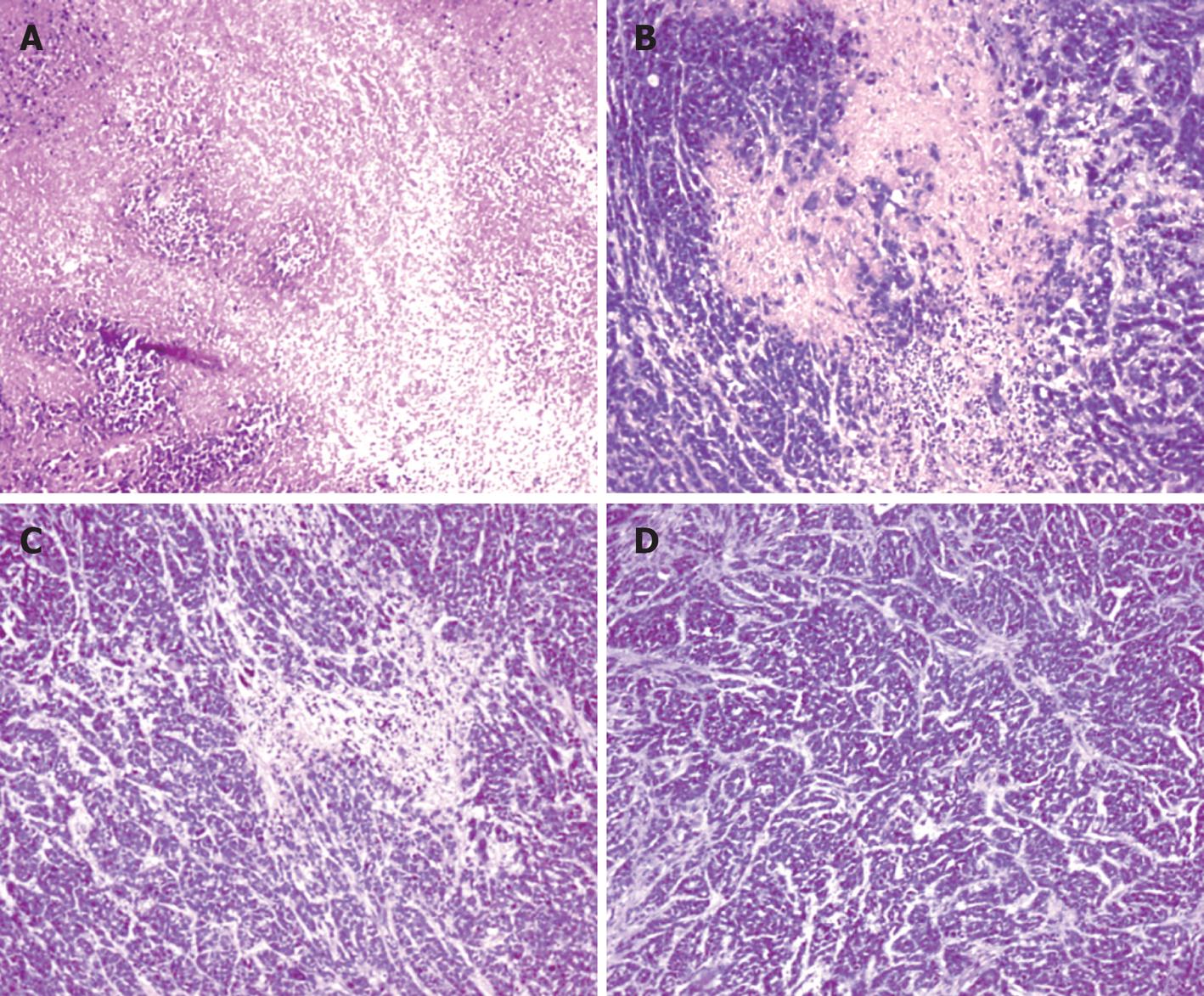
Figure 6 Tumor tissue 28 d after attacked by EC109 showing big necrosis in ED group (A), lamella necrosis in D-group (B), mottling necrosis in E-group (C) and unobvious necrosis in P-group (D) (HE, 10 × 10).
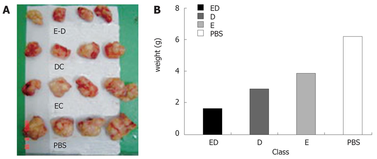
Figure 7 Tumor size (A) and weight (B) in therapeutic group after treatment of EC109-DC, DC, inactivated EC109 and PBS.
Figure 8 Tumor tissue 28 d after attacked by EC109 showing slight necrosis (A), lamella necrosis (B) and mottling necrosis (C) in therapeutic group (HE, 10 × 10) .
-
Citation: Guo GH, Chen SZ, Yu J, Zhang J, Luo LL, Xie LH, Su ZJ, Dong HM, Xu H, Wu LB.
In vivo anti-tumor effect of hybrid vaccine of dendritic cells and esophageal carcinoma cells on esophageal carcinoma cell line 109 in mice with severe combined immune deficiency. World J Gastroenterol 2008; 14(8): 1167-1174 - URL: https://www.wjgnet.com/1007-9327/full/v14/i8/1167.htm
- DOI: https://dx.doi.org/10.3748/wjg.14.1167


