Copyright
©2008 The WJG Press and Baishideng.
World J Gastroenterol. Feb 14, 2008; 14(6): 969-973
Published online Feb 14, 2008. doi: 10.3748/wjg.14.969
Published online Feb 14, 2008. doi: 10.3748/wjg.14.969
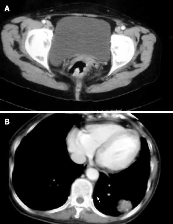
Figure 1 CT scan of the pelvic (A) and chest (B) showing the thickening wall of rectum and an irregularly shaped solid mass (3.
5 cm in diameter) in the lower lobe of the left lung of case 1.
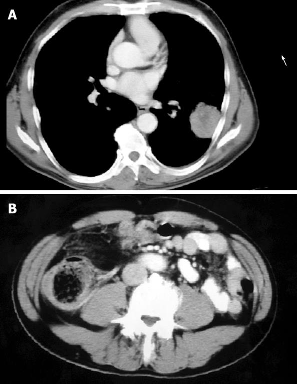
Figure 2 CT scan of the chest (A) and abdomen (B) showing an anomalous round solid mass in the lower lobe of the left lung and an intraluminal mass in the ascending colon with an irregular and stenotic lumen in case 2.
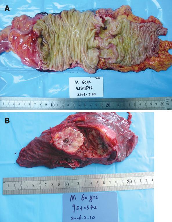
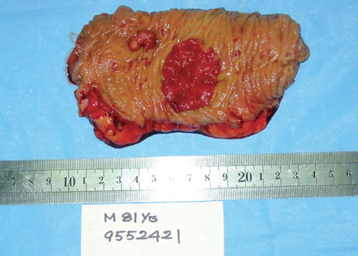
Figure 3 Surgical specimens of the colon cancer (A) and ascending cancer (B) from case three.
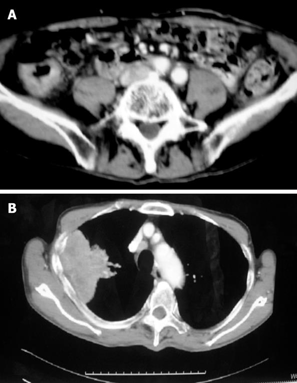
Figure 4 CT scan of the abdomen (A) and chest (B) showing a mass in the ascending colon and an irregularly shaped mass in the upper lobe of the right lung.
Figure 5 Surgical resection specimens of the ascending colon.
- Citation: Peng YF, Gu J. Synchronous colorectal and lung cancer: Report of three cases. World J Gastroenterol 2008; 14(6): 969-973
- URL: https://www.wjgnet.com/1007-9327/full/v14/i6/969.htm
- DOI: https://dx.doi.org/10.3748/wjg.14.969


