Copyright
©2008 The WJG Press and Baishideng.
World J Gastroenterol. Feb 14, 2008; 14(6): 954-959
Published online Feb 14, 2008. doi: 10.3748/wjg.14.954
Published online Feb 14, 2008. doi: 10.3748/wjg.14.954
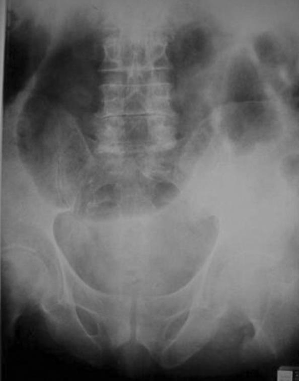
Figure 1 Supine radiograph with no preparation showing massive colonic distention especially the transverse colon, but without air-fluid levels.
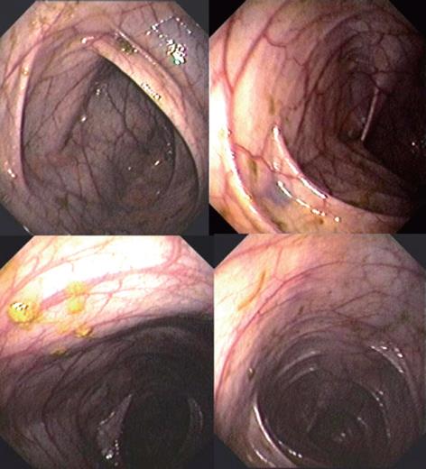
Figure 2 Colonoscopy showing huge colonic distension involving all segments, with a diameter of the rectosigmoid and descending colon greater than 12 cm, and that of the ascending colon and cecum greater than 16-18 cm.
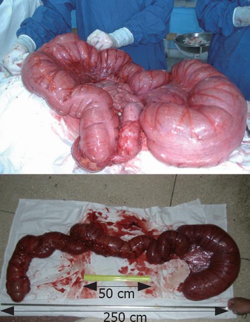
Figure 3 Laparotomy showing massive distension of the whole colon with no perforation or volvulus.
Total colectomy removed a completely atonic colon which was 2.5 m in length and 25 cm in diameter. Ileoanal pouch was made subsequently.
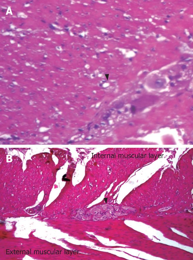
Figure 4 Full-thickness serial biopsies stained with haematoxylin-eosin (× 100) from the removed colon showing no ganglionic cells in the last 7 cm of the rectum.
Marked depletion of ganglionic cells (arrow) and hypertrophy of the external muscular layer were identified in sigmoid (A) and descending colon (B).
- Citation: Georgescu EF, Vasile I, Ionescu R. Intestinal pseudo-obstruction: An uncommon condition with heterogeneous etiology and unpredictable outcome. World J Gastroenterol 2008; 14(6): 954-959
- URL: https://www.wjgnet.com/1007-9327/full/v14/i6/954.htm
- DOI: https://dx.doi.org/10.3748/wjg.14.954


