Copyright
©2008 The WJG Press and Baishideng.
World J Gastroenterol. Dec 21, 2008; 14(47): 7267-7270
Published online Dec 21, 2008. doi: 10.3748/wjg.14.7267
Published online Dec 21, 2008. doi: 10.3748/wjg.14.7267
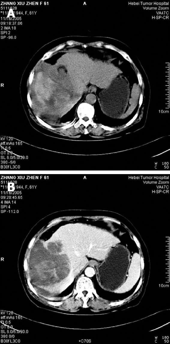
Figure 1 CT scan befor surgery showing a large mass (A) and a well-circumscribed and lowly-attenuated mass (B) in the right liver lobe.
Figure 2 UESL.
A: Atypical spindle cells mixed with stellated cells (HE, × 200); B: Normal bile ducts in the tumor (HE, × 200); C: Normal hepatic cells in the tumor (HE, × 200); D: Giant cells containing eosinophilic hyaline globules in the cytoplasm (HE, × 400).
Figure 3 Immunohistochemical expression of UESL.
A: Strong reactivity for vimentin in spindle and polygonal or round cells (streptavidin-biotin method, × 200); B: Tumor cells containing eosinophilic hyaline globules with positive PAS.
- Citation: Ma L, Liu YP, Geng CZ, Tian ZH, Wu GX, Wang XL. Undifferentiated embryonal sarcoma of liver in an old female: Case report and review of the literature. World J Gastroenterol 2008; 14(47): 7267-7270
- URL: https://www.wjgnet.com/1007-9327/full/v14/i47/7267.htm
- DOI: https://dx.doi.org/10.3748/wjg.14.7267


