Copyright
©2008 The WJG Press and Baishideng.
World J Gastroenterol. Oct 28, 2008; 14(40): 6249-6253
Published online Oct 28, 2008. doi: 10.3748/wjg.14.6249
Published online Oct 28, 2008. doi: 10.3748/wjg.14.6249
Figure 1 The location of MK in HepG2 cells nucleolus using immunogold labeling electron microscopy.
HepG2 cells labelled without MK antibody were performed as the control. No immunogold particles of MK were seen (A). The MK protein (arrow) mostly localized to the DFC, GC and the region between FC and DFC (B, C) [scale bar represents 0.5 μm (A, B) and 1 μm (C)].
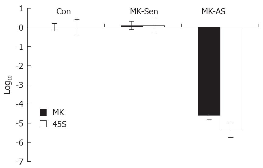
Figure 2 45S rRNA transcription could be regulated by endogenous MK level.
It was shown that 45S rRNA transcription was decreased significantly in response to downregulation of MK expression, through real-time PCR analysis (P < 0.05).
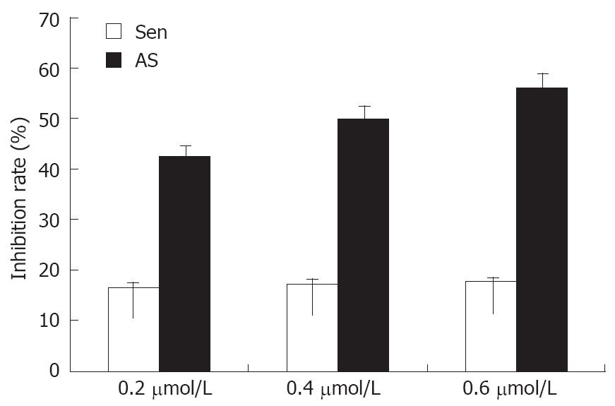
Figure 3 Effect of MK on proliferation of HepG2 cells.
HepG2 cells were transfected with 0.2, 0.4 and 0.6 μmol/L MK-As or MK-Sen for 24 h, and were analyzed by MTT assay. Data show that HepG2 cell proliferation and growth were inhibited by downregulating the MK expression with antisense MK transfection (P < 0.05).
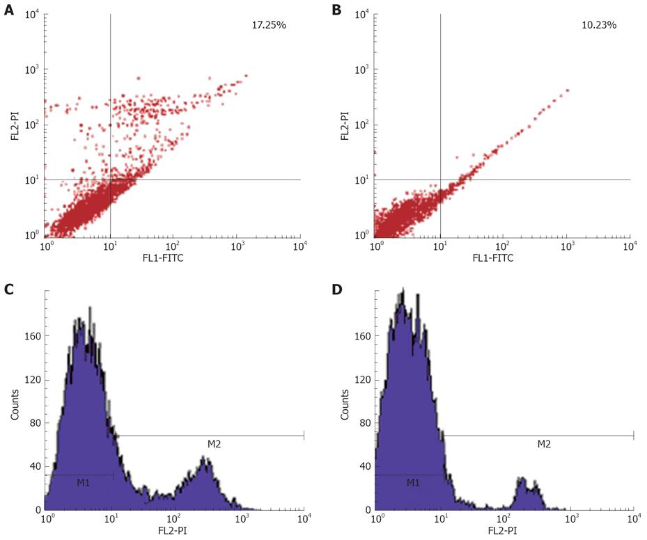
Figure 4 Exogenous MK mediates its anti-apoptotic activity.
HepG2 cells are induced to apoptosis by 10-6 mol/L adriamycin for 20 h. It shows that about 17.25% of cells enter apoptosis (A, C), while 500 ng/mL exogenous MK showed its anti-apoptotic of activity (B, D).
- Citation: Dai LC, Shao JZ, Min LS, Xiao YT, Xiang LX, Ma ZH. Midkine accumulated in nucleolus of HepG2 cells involved in rRNA transcription. World J Gastroenterol 2008; 14(40): 6249-6253
- URL: https://www.wjgnet.com/1007-9327/full/v14/i40/6249.htm
- DOI: https://dx.doi.org/10.3748/wjg.14.6249


