Copyright
©2008 The WJG Press and Baishideng.
World J Gastroenterol. Sep 21, 2008; 14(35): 5474-5477
Published online Sep 21, 2008. doi: 10.3748/wjg.14.5474
Published online Sep 21, 2008. doi: 10.3748/wjg.14.5474
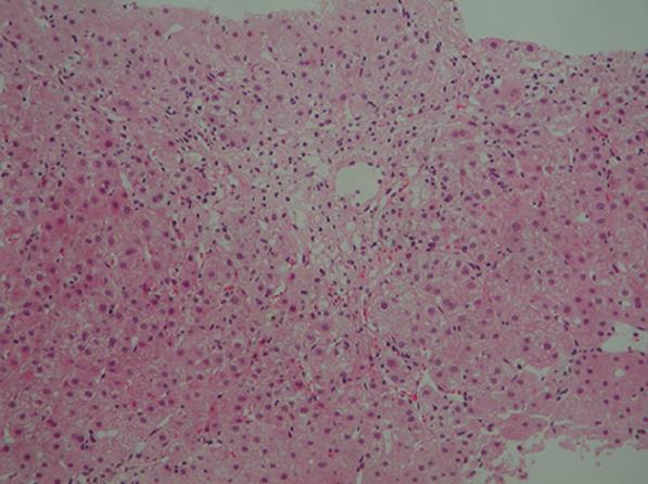
Figure 1 Histological examination of a liver biopsy specimen showing bridging perivenular necrosis and infiltration of inflammatory cells including eosinophils (hematoxylin-eosin staining × 100).
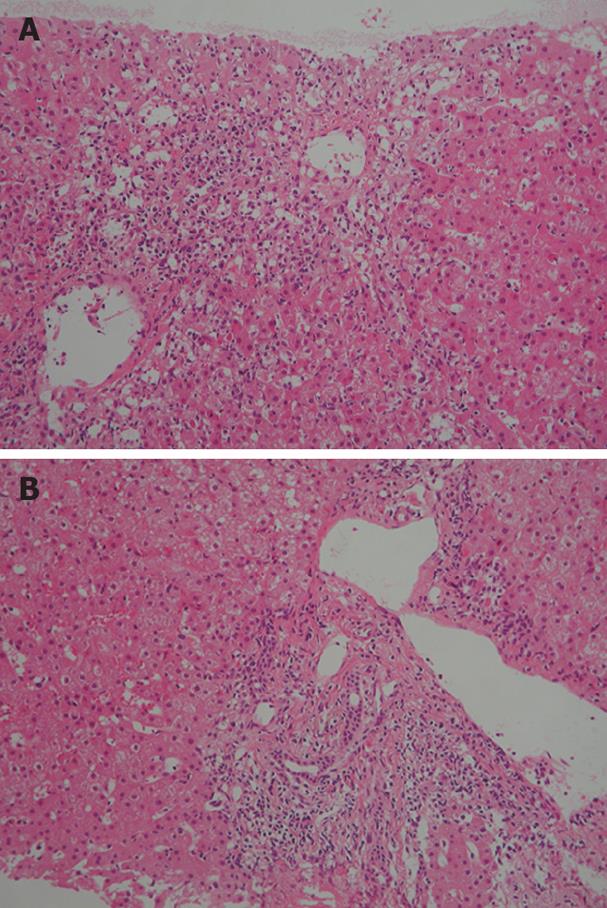
Figure 2 Histological examination of a liver biopsy specimen showing bridging perivenular necrosis (A) and interface hepatitis (B) (HE staining × 100).
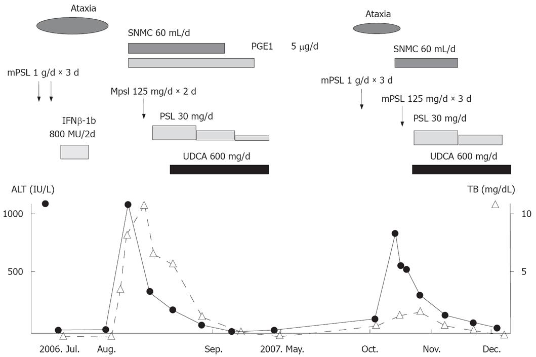
Figure 3 Clinical course of the disease.
SNMC: Stronger neo-minophagen C, PGE1: Prostaglandin, mPSL: Methylprednisolone, PSL: Prednisolone, IFN-β: Interferon β.
- Citation: Takahashi A, Kanno Y, Takahashi Y, Sakamoto N, Monoe K, Saito H, Abe K, Yokokawa J, Irisawa A, Ohira H. Development of autoimmune hepatitis type 1 after pulsed methylprednisolone therapy for multiple sclerosis: A case report. World J Gastroenterol 2008; 14(35): 5474-5477
- URL: https://www.wjgnet.com/1007-9327/full/v14/i35/5474.htm
- DOI: https://dx.doi.org/10.3748/wjg.14.5474


