Copyright
©2008 The WJG Press and Baishideng.
World J Gastroenterol. Aug 28, 2008; 14(32): 5000-5007
Published online Aug 28, 2008. doi: 10.3748/wjg.14.5000
Published online Aug 28, 2008. doi: 10.3748/wjg.14.5000
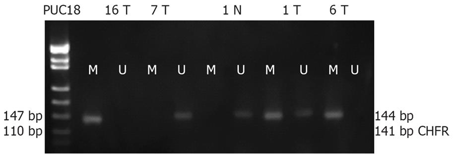
Figure 1 Representative results of MSPCR in human GC tissue samples.
Lanes U and M: Products derived from unmethylated and methylated alleles, respectively. Methylation of the CHFR gene was detected in 16 and 6 poorly-differentiated adenocarcinoma tissue samples and 1 mucinous adenocarcinoma tissue sample, while unmethylation of the CHFR gene was detected in 7 well-differentiated adenocarcinoma tissue samples. PUC18: Marker; N: Normal gastric mucosa corresponding to tumors; T: Gastric cancer.
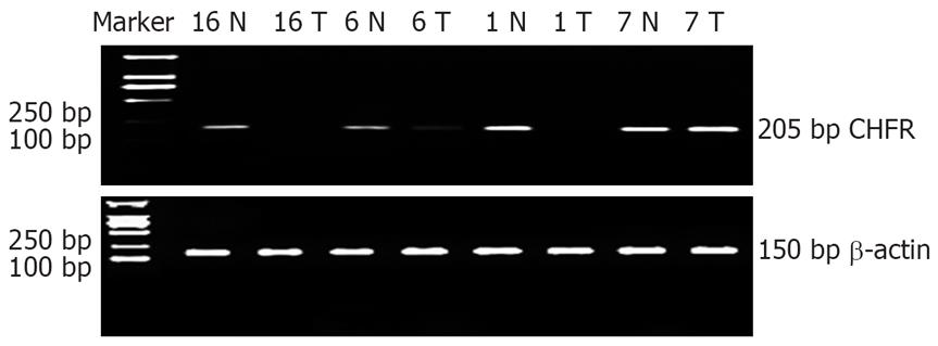
Figure 2 Representative results of RT-PCR in human GC tissue samples.
β-actin was used as an internal control. CHFR mRNA expression level in 16 and 6 poorly differentiated adenocarcinoma tissue samples and 1 mucinous adenocarcinoma tissue sample was significantly lower than that in paired normal gastric mucosa samples from the same patients, while no difference was found in 7 well-differentiated adenocarcinoma tissue samples. Marker, D2000: Marker; N: Normal gastric mucosa corresponding to tumors; T: Gastric cancer.
Figure 3 Representative results of Western blot in human GC tissue samples.
β-tubulin was used as an internal control. CHFR protein expression level in GS tissue samples was significantly lower than that in paired normal gastric mucosa samples. Aberrant methylation of CHFR and down-regulation or loss of CHFR mRNA expression were detected in 16 poorly differentiated adenocarcinoma tissue samples and 1 mucinous adenocarcinoma tissue sample, while positive protein expression of CHFR was detected in 9 moderately-differentiated adenocarcinoma tissue samples without CHFR methylation. N: Normal gastric mucosa corresponding to tumors; T: Gastric cancer.
Figure 4 Immunohistochemical staining for CHFR protein expression in GC tissue samples and normal gastric mucosa samples.
Positive expression of CHFR in normal gastric mucosa tissue samples (A), in well-differentiated adenocarcinoma tissue samples (B), and in signet-ring cell carcinoma tissue samples (C).
- Citation: Gao YJ, Xin Y, Zhang JJ, Zhou J. Mechanism and pathobiologic implications of CHFR promoter methylation in gastric carcinoma. World J Gastroenterol 2008; 14(32): 5000-5007
- URL: https://www.wjgnet.com/1007-9327/full/v14/i32/5000.htm
- DOI: https://dx.doi.org/10.3748/wjg.14.5000


