Copyright
©2008 The WJG Press and Baishideng.
World J Gastroenterol. Aug 14, 2008; 14(30): 4838-4840
Published online Aug 14, 2008. doi: 10.3748/wjg.14.4838
Published online Aug 14, 2008. doi: 10.3748/wjg.14.4838
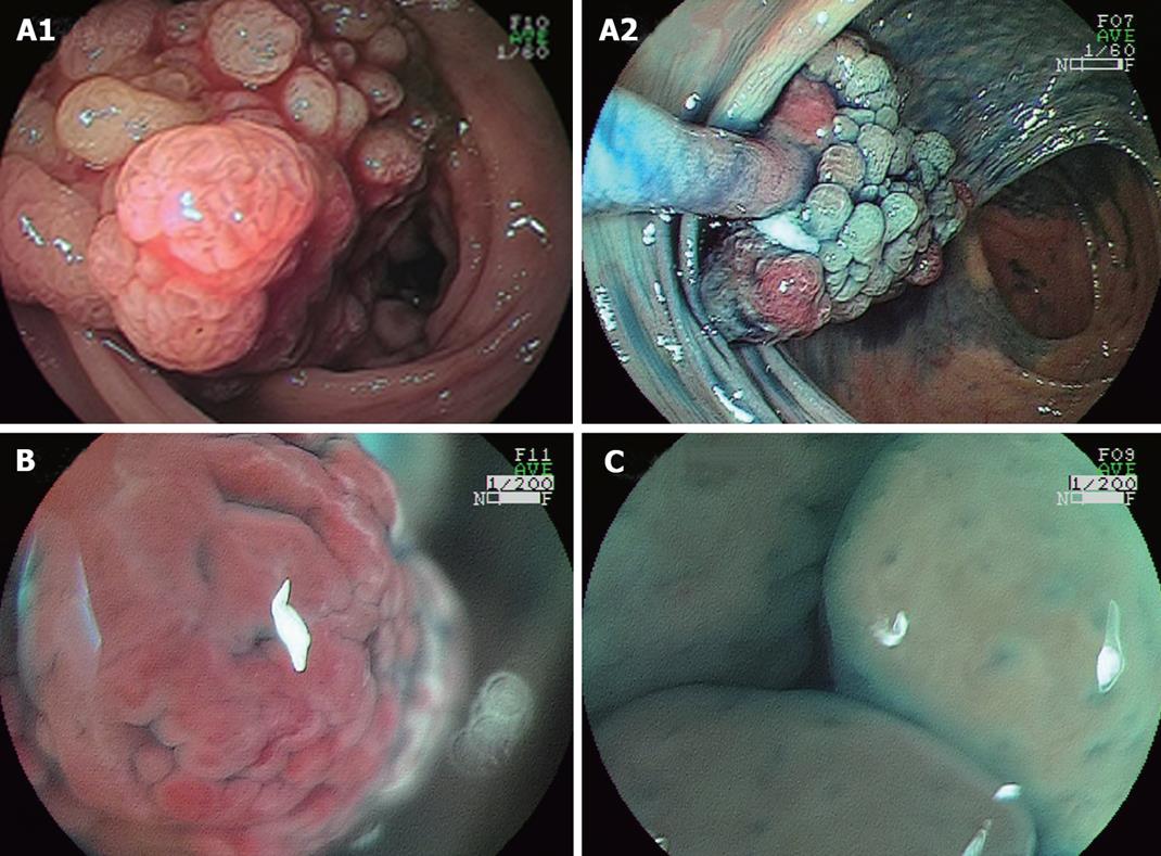
Figure 1 Endoscopy showing a lobulated, peduncular polyp with bleeding, about 40 mm in diameter, in the ascending colon with both red and white components and a mosaic pattern observed in the polyp (A), magnifying endoscopy revealing a rugged surface of red component without normal mucosal structure (× 50) (B), and aggregated smooth nodules with enlarged round or oval crypt openings in the white component (× 50) (C).
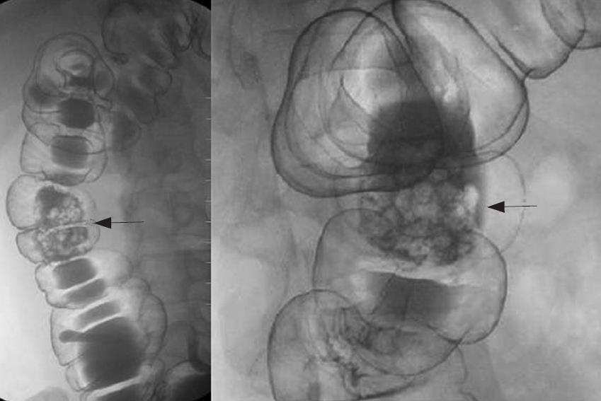
Figure 2 Double-contrast radiograph of the ascending colon showing an about 40 mm lobulated peduncular polyp (arrow).
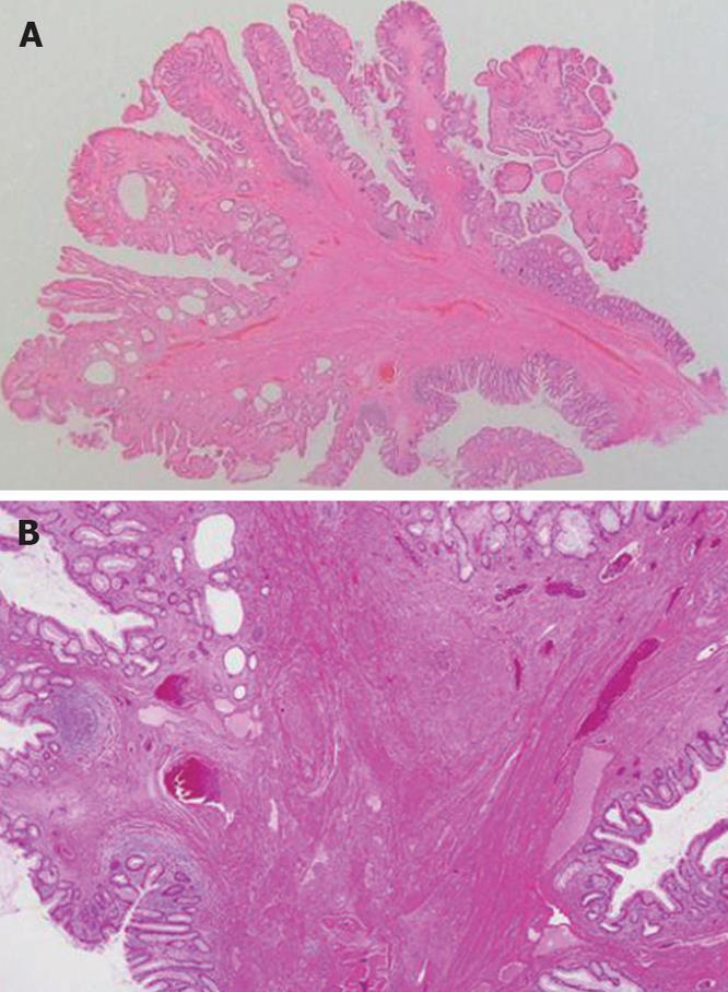
Figure 3 Microscopy of the polypectomy specimen showing a stalked polyp containing numerous cystically dilated glands on the cross section under lower-power view (HE, × 4) (A) and inflammatory granulation tissue in the lamina propria mucosae and proliferation of smooth muscle (HE, × 50) (B).
- Citation: Kanzaki H, Hirasaki S, Okuda M, Kudo K, Suzuki S. Lobulated inflammatory myoglandular polyp in the ascending colon observed by magnifying endoscopy and treated with endoscopic polypectomy. World J Gastroenterol 2008; 14(30): 4838-4840
- URL: https://www.wjgnet.com/1007-9327/full/v14/i30/4838.htm
- DOI: https://dx.doi.org/10.3748/wjg.14.4838


