Copyright
©2008 The WJG Press and Baishideng.
World J Gastroenterol. Jan 21, 2008; 14(3): 484-486
Published online Jan 21, 2008. doi: 10.3748/wjg.14.484
Published online Jan 21, 2008. doi: 10.3748/wjg.14.484
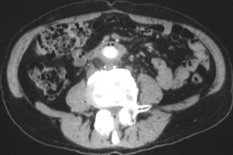
Figure 1 CT scan showing an area with the characteristics of inflammatory tissue including air bubbles between the duodenum and aortic-bi-femoral prosthesis adherent to the third duodenal portion.
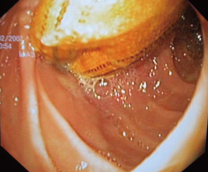
Figure 2 Esophago-gastro-duodenoscopy showing the aortic prosthesis crossing the third segment of the duodenal wall occluding the intestinal lumen.
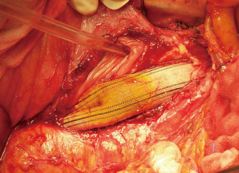
Figure 3 Isolated prosthesis in the context of eroded duodenal wall during laparotomy (the tube indicates the proximal duodenal lumen).
- Citation: Geraci G, Pisello F, Volsi FL, Facella T, Platia L, Modica G, Sciumè C. Secondary aortoduodenal fistula. World J Gastroenterol 2008; 14(3): 484-486
- URL: https://www.wjgnet.com/1007-9327/full/v14/i3/484.htm
- DOI: https://dx.doi.org/10.3748/wjg.14.484


