Copyright
©2008 The WJG Press and Baishideng.
World J Gastroenterol. Aug 7, 2008; 14(29): 4697-4700
Published online Aug 7, 2008. doi: 10.3748/wjg.14.4697
Published online Aug 7, 2008. doi: 10.3748/wjg.14.4697
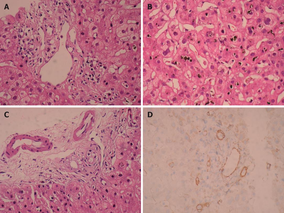
Figure 1 Moderate predominantly lymphocytic mixed portal infiltration and absence of interlobular bile duct destruction (HE, × 400) (A), intralobular severe canalicular cholestasis (HE, × 400) (B), marked cytoplasmic vacuolization in cholangiolar epithelium (HE, × 400) (C), and absence of bile ducts by cytokeratin 19 immunohistochemical staining (IHC, × 400) (D), observed in our patient.
Figure 2 ALT and ALP (A) and total biluribin (B) levels during and after treatment in our case.
- Citation: Okan G, Yaylaci S, Peker O, Kaymakoglu S, Saruc M. Vanishing bile duct and Stevens-Johnson syndrome associated with ciprofloxacin treated with tacrolimus. World J Gastroenterol 2008; 14(29): 4697-4700
- URL: https://www.wjgnet.com/1007-9327/full/v14/i29/4697.htm
- DOI: https://dx.doi.org/10.3748/wjg.14.4697


