Copyright
©2008 The WJG Press and Baishideng.
World J Gastroenterol. Jul 21, 2008; 14(27): 4296-4299
Published online Jul 21, 2008. doi: 10.3748/wjg.14.4296
Published online Jul 21, 2008. doi: 10.3748/wjg.14.4296
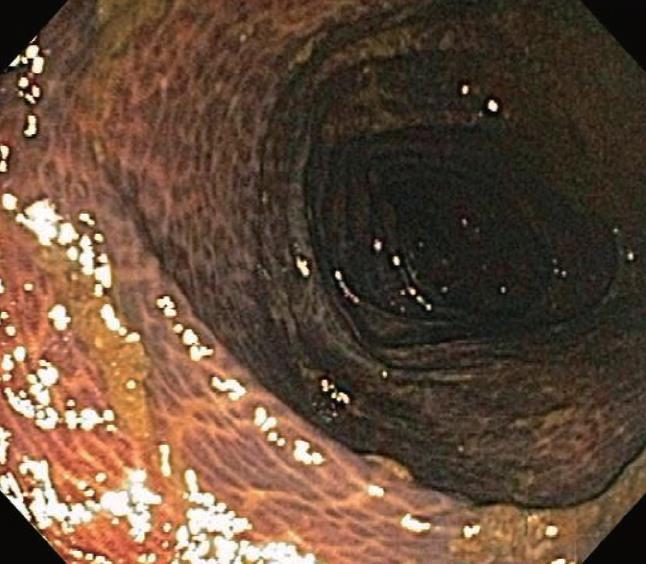
Figure 1 Typical alligator or snake-skin appearance of melanosis coli.
Despite routine colon preparation, residual fecal debris is common, likely reflecting reduced colonic propulsive activity.
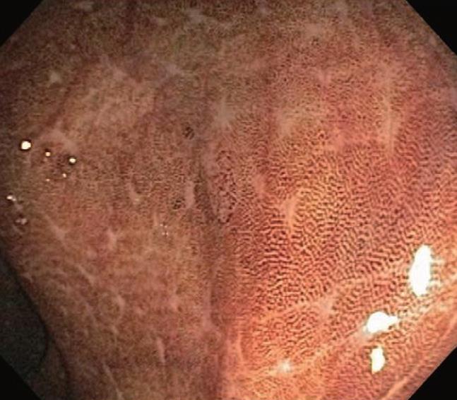
Figure 2 Rectosigmoid mucosa illustrating focal areas of patchy intense pigmentation as well as focal areas of absent pigment, the latter reflecting the presence of normal mucosal aggregates of lymphoid cells (so-called “starry sky” appearance of melanosis coli).
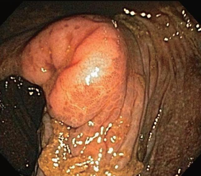
Figure 3 Ileal and cecal mucosa in melanosis coli.
Melanosis is generally confined to the cecal mucosa although there is a very limited area of transition into ileal mucosa that is not pigmented.
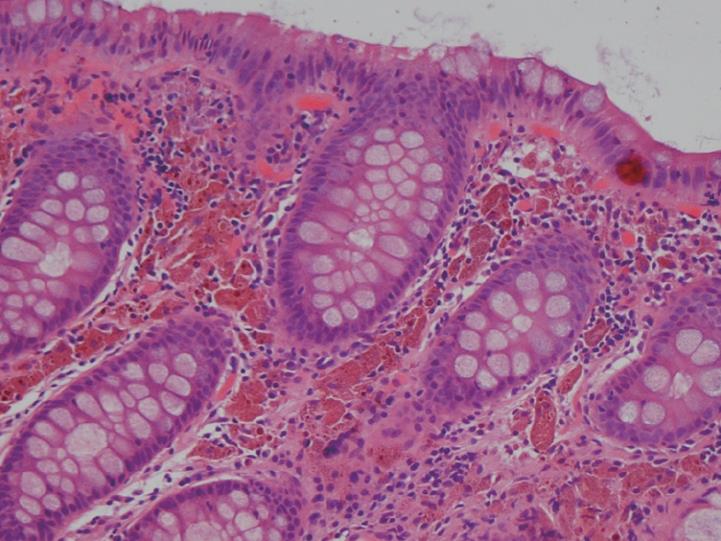
Figure 4 Photomicrograph showing typical pigment granule laden macrophages in the lamina propria.
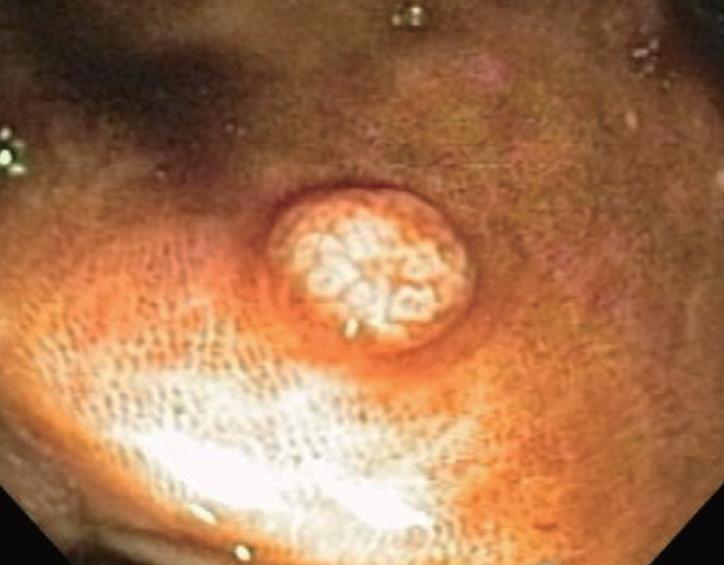
Figure 5 Easily visualized small sessile polypoid lesion in ascending colon with adjacent background pigmented mucosa typical of melanosis coli.
Resected specimen confirmed absence of pigmented macrophages in the body of the resected adenoma.
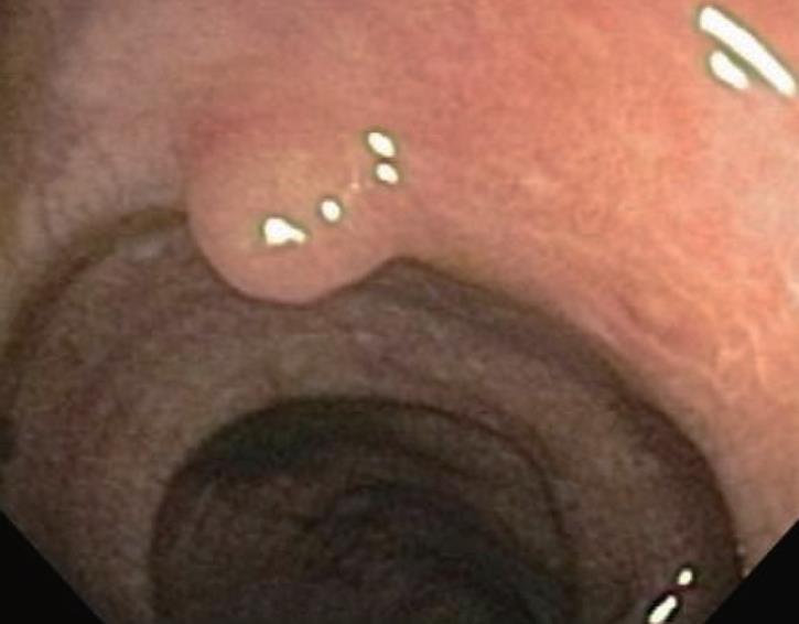
Figure 6 Small pigmented polypoid lesion similar to background pigmented colonic mucosa.
Resected specimen revealed a submucosal leiomyoma along with pigmented macrophages in the overlying colonic mucosa due to melanosis coli.
- Citation: Freeman HJ. “Melanosis” in the small and large intestine. World J Gastroenterol 2008; 14(27): 4296-4299
- URL: https://www.wjgnet.com/1007-9327/full/v14/i27/4296.htm
- DOI: https://dx.doi.org/10.3748/wjg.14.4296


