Copyright
©2008 The WJG Press and Baishideng.
World J Gastroenterol. Jun 21, 2008; 14(23): 3681-3692
Published online Jun 21, 2008. doi: 10.3748/wjg.14.3681
Published online Jun 21, 2008. doi: 10.3748/wjg.14.3681
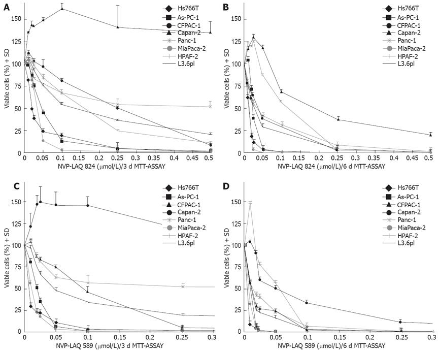
Figure 1 In vitro treatment of pancreatic cancer with NVP-LAQ824 and NVP-LBH589 (MTT assay).
A: 3-d incubation with NVP-LAQ824 (n = 3); B: 6-d incubation with NVP-LAQ824 (n = 3); C: 3-d incubation with NVP-LBH589 (n = 3); D: 6-d incubation with NVP-LBH589 (n = 3).
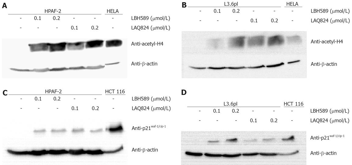
Figure 2 Mechanism of drug action after in vitro treatment with NVP-LAQ824 and NVP-LBH589 for 24 h.
A and B: Acetylation of histone H4. Protein extracts from HELA cells that were treated with 5 mmol/L sodium butyrate served as positive controls; C and D: p21WAF-1/CIP-1 expression. Cell lysate from HCT 116 colon cancer cells served as positive control; A-D: Staining with β-actin antibody confirmed equal protein loading.
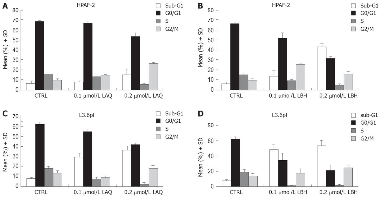
Figure 3 Cell cycle analysis.
A: Treatment of cell line HPAF-2 with 0.1 or 0.2 &mgr;mol/L NVP-LAQ824 for 72 h (n = 3); B: Treatment of cell line HPAF-2 with 0.1 or 0.2 &mgr;mol/L NVP-LBH589 for 72 h (n = 3); C: Treatment of cell line L3.6pl with 0.1 or 0.2 &mgr;mol/L NVP-LAQ824 for 72 h (n = 3); D: Treatment of cell line L3.6pl with 0.1 or 0.2 &mgr;mol/L NVP-LBH589 for 72 h (n = 3).
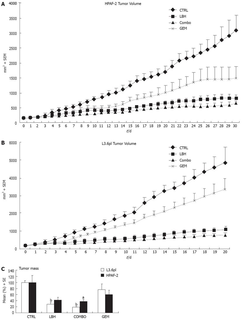
Figure 4 In vivo treatment with NVP-LBH589 + gemcitabine in chimeric mice.
A: Effect on tumor volume of HPAF-2 cells; B: Effect on tumor volume of L3.6pl cells; C: Effect on tumor mass (aP < 0.05, COMBO vs control; bP < 0.01, NVP-LBH589 or COMBO vs control).
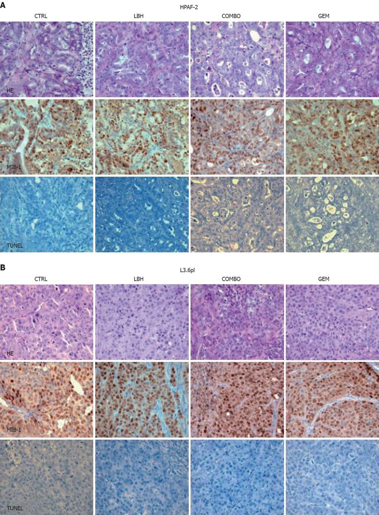
Figure 5 Hematoxylin-eosin (HE), MIB-1 (proliferation marker) and TUNEL (apoptosis marker) staining of mouse tumors (SABC, x 40).
A: Cell line HPAF-2; B: Cell line L3.6pl.
- Citation: Haefner M, Bluethner T, Niederhagen M, Moebius C, Wittekind C, Mossner J, Caca K, Wiedmann M. Experimental treatment of pancreatic cancer with two novel histone deacetylase inhibitors. World J Gastroenterol 2008; 14(23): 3681-3692
- URL: https://www.wjgnet.com/1007-9327/full/v14/i23/3681.htm
- DOI: https://dx.doi.org/10.3748/wjg.14.3681


