Copyright
©2008 The WJG Press and Baishideng.
World J Gastroenterol. Jun 7, 2008; 14(21): 3430-3434
Published online Jun 7, 2008. doi: 10.3748/wjg.14.3430
Published online Jun 7, 2008. doi: 10.3748/wjg.14.3430
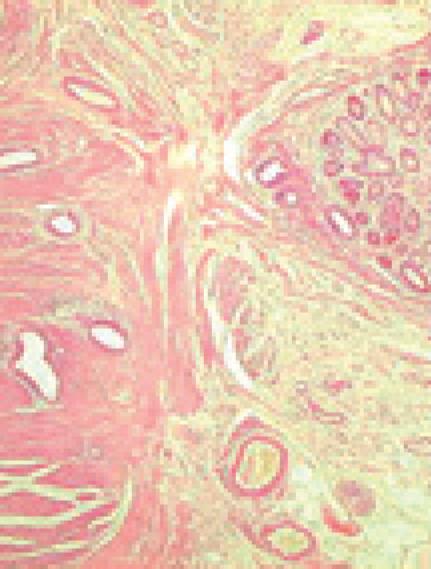
Figure 1 Histology of ileal wall showing endometrial tissue in the muscular layer, with foci of mucosal involvement.
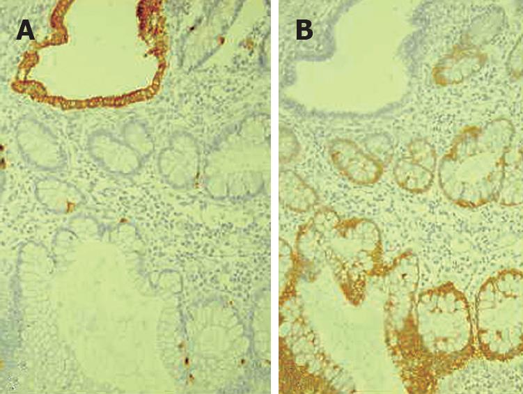
Figure 2 Histopathology showing CK20 immunostaining of intestinal epithelium (A) and CK 7 immunostaining of endometrioid glands (B).
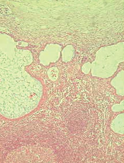
Figure 3 Endometriosis involving lymph nodes with a cystic glandular pattern.
- Citation: Ceglie AD, Bilardi C, Blanchi S, Picasso M, Muzio MD, Trimarchi A, Conio M. Acute small bowel obstruction caused by endometriosis: A case report and review of the literature. World J Gastroenterol 2008; 14(21): 3430-3434
- URL: https://www.wjgnet.com/1007-9327/full/v14/i21/3430.htm
- DOI: https://dx.doi.org/10.3748/wjg.14.3430


