Copyright
©2008 The WJG Press and Baishideng.
World J Gastroenterol. Jun 7, 2008; 14(21): 3410-3415
Published online Jun 7, 2008. doi: 10.3748/wjg.14.3410
Published online Jun 7, 2008. doi: 10.3748/wjg.14.3410
Figure 1 The micrographs of light microscope stained with haematoxylin and eosin.
Micrograph (A) illustrates the typical structure of villi; (B) blunting of the villi (arrow head) and subepithelial edema (asterisk); (C) existing subepithelial edema (asterisk) and the total mucosal thickness (arrow).
Figure 2 These transmission electron microscope (TEM) micrographs illustrate the main ultrastructural features of enterocytes, the absorptive cells of the ileum.
Micrograph (A) shows the regular structure of microvilli (Mv) and the three components of junctional complex at the luminal end of the lateral plasma membrane, zonula occludens (ZO), zonula adherens (ZA) and macula adherens (MA); Micrograph (B) shows the disintegration of the zonula occludens and disordered structure of junctional complexes (arrows). The lipid droplets (L) and phagosomes (P) in the cytoplasm, rough endoplasmic reticulum (rER), swollen mitochondria (M) with ballooned cristae (Bc) and apical surface edema (asterisk) viewed; Micrograph (C) illustrates the regular structure of microvilli (Mv) and junctional complexes (ZO, ZA, MA).
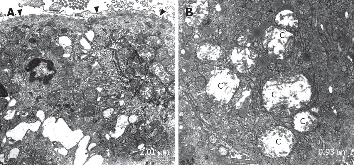
Figure 3 Micrograph (A) shows the disintegration of the zonula occludens (arrow), apical surface edema (asterisk) and the desquamation of the epithelial tissue (arrow head); Micrograph (B) illustrates the swollen mitochondria with cavitations of matrix (C).
- Citation: Gencay C, Kilicoglu SS, Kismet K, Kilicoglu B, Erel S, Muratoglu S, Sunay AE, Erdemli E, Akkus MA. Effect of honey on bacterial translocation and intestinal morphology in obstructive jaundice. World J Gastroenterol 2008; 14(21): 3410-3415
- URL: https://www.wjgnet.com/1007-9327/full/v14/i21/3410.htm
- DOI: https://dx.doi.org/10.3748/wjg.14.3410


