Copyright
©2008 The WJG Press and Baishideng.
World J Gastroenterol. Jun 7, 2008; 14(21): 3410-3415
Published online Jun 7, 2008. doi: 10.3748/wjg.14.3410
Published online Jun 7, 2008. doi: 10.3748/wjg.14.3410
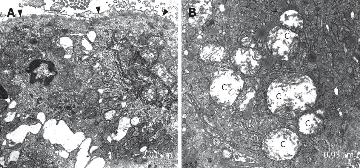
Figure 3 Micrograph (A) shows the disintegration of the zonula occludens (arrow), apical surface edema (asterisk) and the desquamation of the epithelial tissue (arrow head); Micrograph (B) illustrates the swollen mitochondria with cavitations of matrix (C).
- Citation: Gencay C, Kilicoglu SS, Kismet K, Kilicoglu B, Erel S, Muratoglu S, Sunay AE, Erdemli E, Akkus MA. Effect of honey on bacterial translocation and intestinal morphology in obstructive jaundice. World J Gastroenterol 2008; 14(21): 3410-3415
- URL: https://www.wjgnet.com/1007-9327/full/v14/i21/3410.htm
- DOI: https://dx.doi.org/10.3748/wjg.14.3410


