Copyright
©2008 The WJG Press and Baishideng.
World J Gastroenterol. Jan 14, 2008; 14(2): 231-235
Published online Jan 14, 2008. doi: 10.3748/wjg.14.231
Published online Jan 14, 2008. doi: 10.3748/wjg.14.231
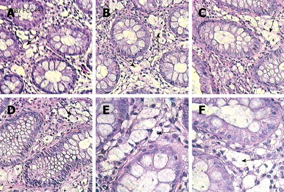
Figure 1 Microscopic features of CCC (A-F) (HE, × 320).
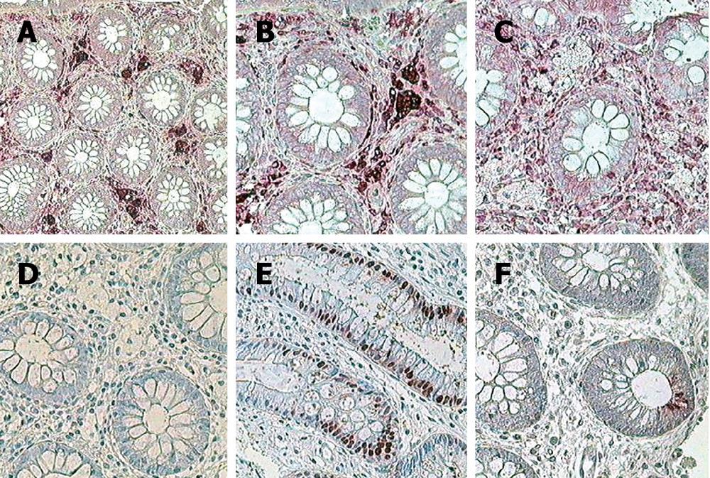
Figure 2 Immunocytochemistry of CCC using monoclonal antibodies and APPAP method showing positive expression of mononuclear phagocytic cell line (A and B), negative T3 and T4 lymphocytes (C), negative neurofilament (D), Normal PCNA expression in cryptal epithelium (E), and normal mitotic activity of positive CD67 (F) (× 320).
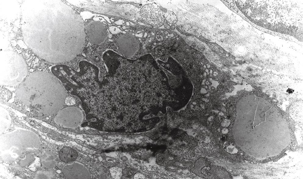
Figure 3 Electron microscopy of clear cells.
Giant clear macrophages contain multiple vesicles varying in size filled with homogenous osmiophylic materials (secondary phagosomes) without any structural components in their cytoplasm (OsO4 staining, × 24 000).
- Citation: Józefczuk J, Wozniewicz BM. Clear cell colitis: A form of microscopic colitis in children. World J Gastroenterol 2008; 14(2): 231-235
- URL: https://www.wjgnet.com/1007-9327/full/v14/i2/231.htm
- DOI: https://dx.doi.org/10.3748/wjg.14.231


