Copyright
©2008 The WJG Press and Baishideng.
World J Gastroenterol. Jan 14, 2008; 14(2): 218-223
Published online Jan 14, 2008. doi: 10.3748/wjg.14.218
Published online Jan 14, 2008. doi: 10.3748/wjg.14.218
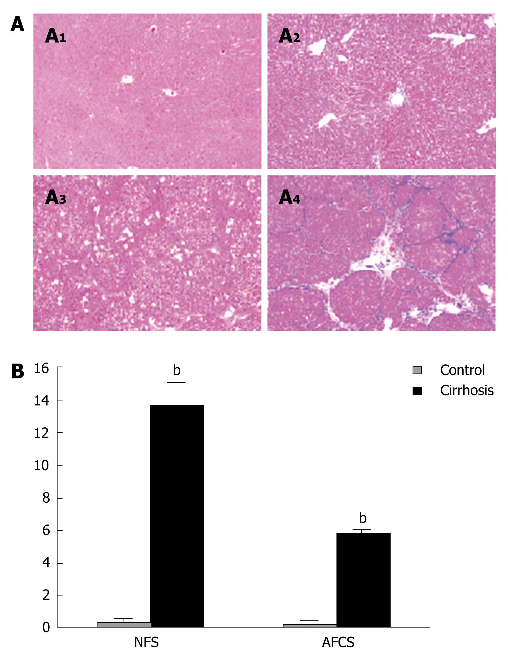
Figure 1 Morphometric analysis of the cirrhotic liver.
A: After 8 wk of CCl4 or vehicle treatment, liver tissue was fixed and stained with hematoxylin/eosin (A1 and A3) as well as Masson's trichrome stain (A2 and A4); B: Necroinflammatory score (NFS) and scores for architectural change, fibrosis and cirrhosis (AFCS) were determined as described in the Methods section. bP < 0.01 vs control.
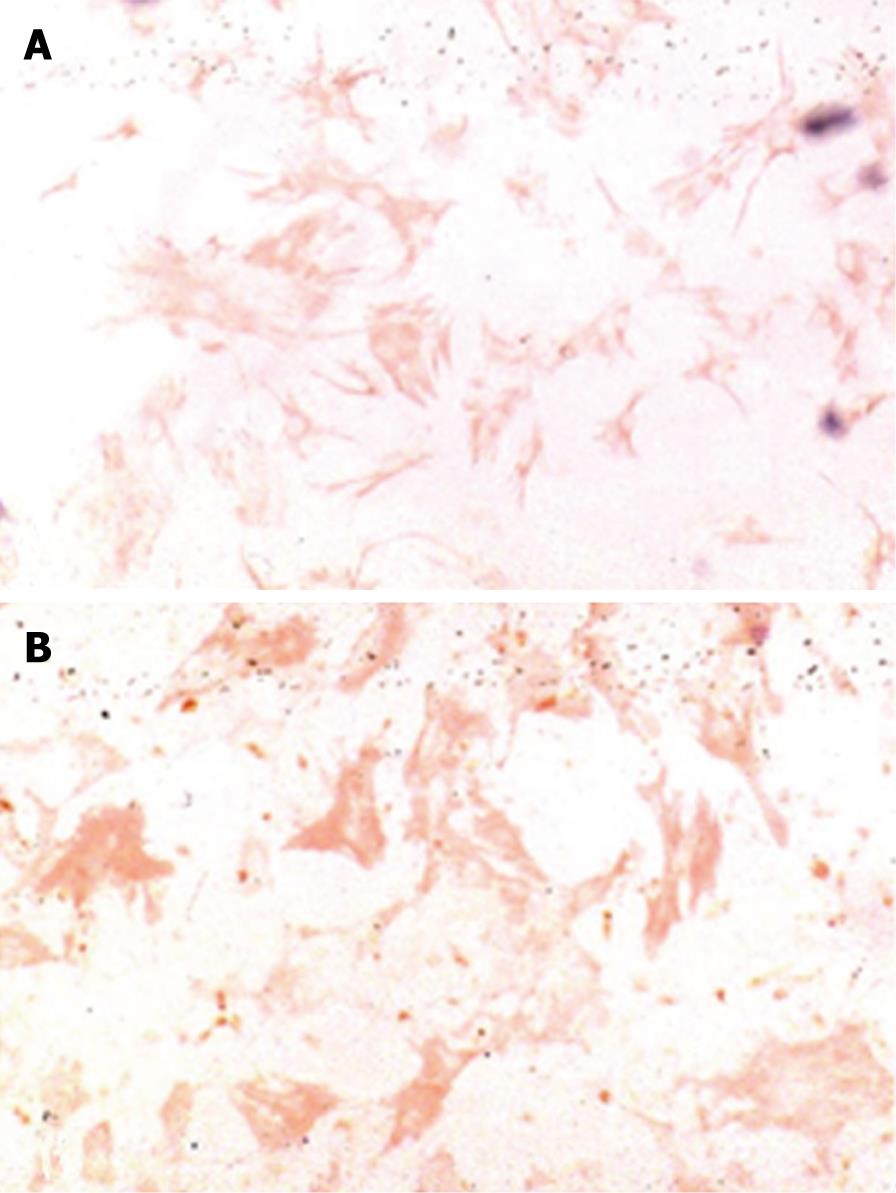
Figure 2 Expression of desmin and aSMA in HSCs freshly isolated from cirrhotic livers.
HSCs from cirrhotic livers show immunostaining for desmin (A) and aSMA (B) (× 400).
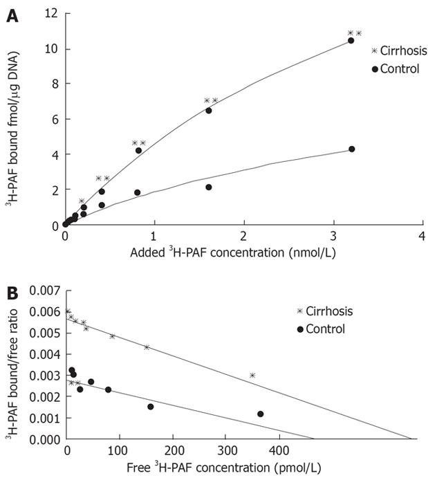
Figure 3 A: Saturation curve of 3H-PAF binding to cultured cirrhotic hepatic stellate cells.
3H-PAF in the concentration between 0.125 and 3.2 nmol/L,in presence or the absence of 5 &mgr;mol/L unlabeled incubated at 25°C for 3 h; B: Scatchard plot analysis of binding of 3H-PAF to cirrhotic hepatic stellate cells. Cirrhosis: Kd: 4.66 nmol/L, Bax: 24.65 ± 1.96 fmol/&mgr;g DNA, R = 0.982; Control: Kd: 3.51 nmol/L, Bax: 5.74 ± 1.55 fmol/&mgr;g DNA, R = 0.93.
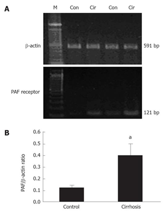
Figure 4 PAF receptor expression in cirrhotic hepatic stellate cells.
RT-PCR of PAF receptor mRNA was performed with cDNA prepared from RNA samples of control and cirrhotic HSCs. Expression of β-actin mRNA was assessed using the same amout of cDNA. A: PCR products of PAF and β-actin from control (Con) and cirrhotic (Cir) rat livers are shown; B: Ratio of the PAF receptor and β-actin mRNA. aP < 0.05 vs control.
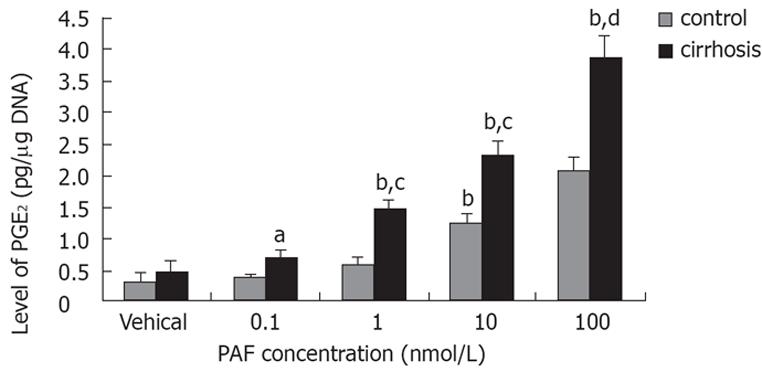
Figure 5 Effect of PAF concentration on PGE2 synthesis in cirrhotic hepatic stellate cells.
Cell were placed in serum-free medium after overnight culture, and were challenged with PAF 24 h later. PGE2 in the medium was measured by ELISA after 15 min incubation. aP < 0.05, bP < 0.01 vs vehical; cP < 0.05, dP < 0.01 vs control.
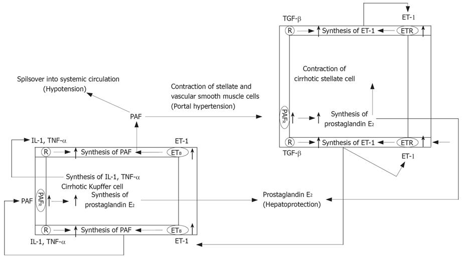
Figure 6 Implication of platelate-activating factor, ET-1, Kupffer cells and hepatic stellates cells in the pathology of liver cirrhosis.
Increased ET-1 released by stellates and endothelial cells in the cirrhosis liver acts on upregulated ETB receptors in Kupffer cells and stimulates the synthesis of PAF. Likewise, autocrine actions of proinflammatory mediator such as IL-1 and TNF-α may also stimulate PAF synthesis by Kupffer cells. PAF then acts on hepatic vascular smooth muscle cells and stellate cells and contributes to portal hypertension by causing their contraction. Its spillover into systemic circulation is likely a cause of hypertension associated with cirrhosis. Autocrine PAF and paracrine ET-1 can act on their respective upregulated receptors in Kupffer cells and stellate cells to cause the synthesis and release of prostaglandin E2, which in turn may act to limit the liver injury.
- Citation: Chen Y, Wang CP, Lu YY, Zhou L, Su SH, Jia HJ, Feng YY, Yang YP. Hepatic stellate cells may be potential effectors of platelet activating factor induced portal hypertension. World J Gastroenterol 2008; 14(2): 218-223
- URL: https://www.wjgnet.com/1007-9327/full/v14/i2/218.htm
- DOI: https://dx.doi.org/10.3748/wjg.14.218


