Copyright
©2008 The WJG Press and Baishideng.
World J Gastroenterol. May 14, 2008; 14(18): 2915-2916
Published online May 14, 2008. doi: 10.3748/wjg.14.2915
Published online May 14, 2008. doi: 10.3748/wjg.14.2915
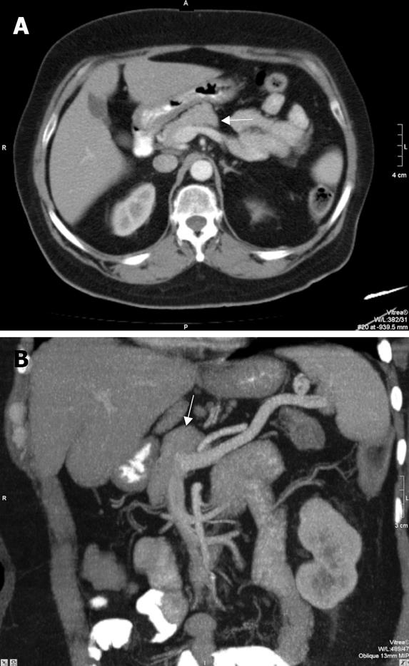
Figure 1 Axial CT (A) and thick-slice coronal oblique MIP CT (B) images show the pancreatic head (arrow) and the absence of the corpus and tail of the pancreas.
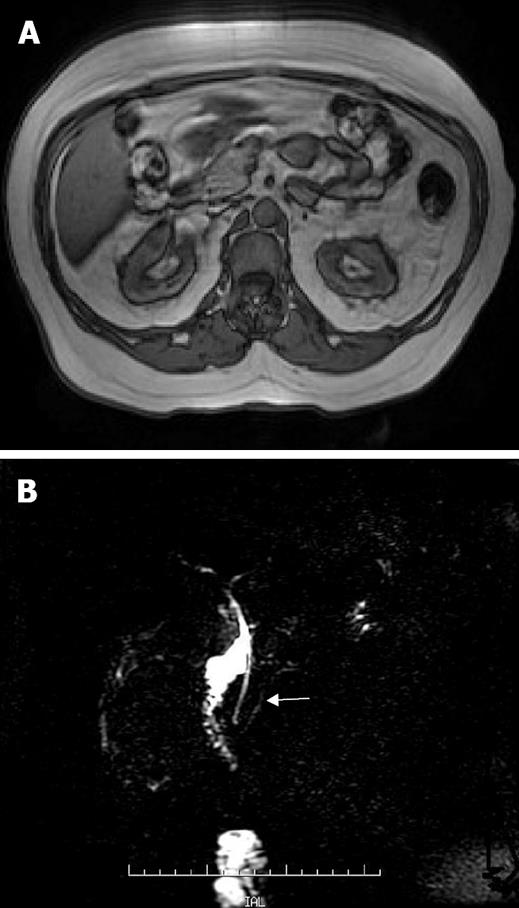
Figure 2 Abdominal axial T1-weighted MR image (A) reveals pancreatic head; MRCP demontrates a short major pancreatic duct (arrow) and dorsal duct is not visualized (B).
- Citation: Pasaoglu L, Vural M, Hatipoglu HG, Tereklioglu G, Koparal S. Agenesis of the dorsal pancreas. World J Gastroenterol 2008; 14(18): 2915-2916
- URL: https://www.wjgnet.com/1007-9327/full/v14/i18/2915.htm
- DOI: https://dx.doi.org/10.3748/wjg.14.2915


