Copyright
©2008 The WJG Press and Baishideng.
World J Gastroenterol. Apr 28, 2008; 14(16): 2612-2614
Published online Apr 28, 2008. doi: 10.3748/wjg.14.2612
Published online Apr 28, 2008. doi: 10.3748/wjg.14.2612
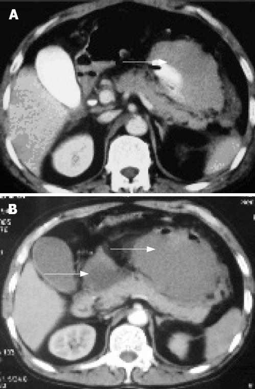
Figure 1 CT scan.
A: Presence of the postperitoneal hematoma (arrow); B: Increase in the size of postperitoneal hematoma and presence of a pseudocyst (arrows).
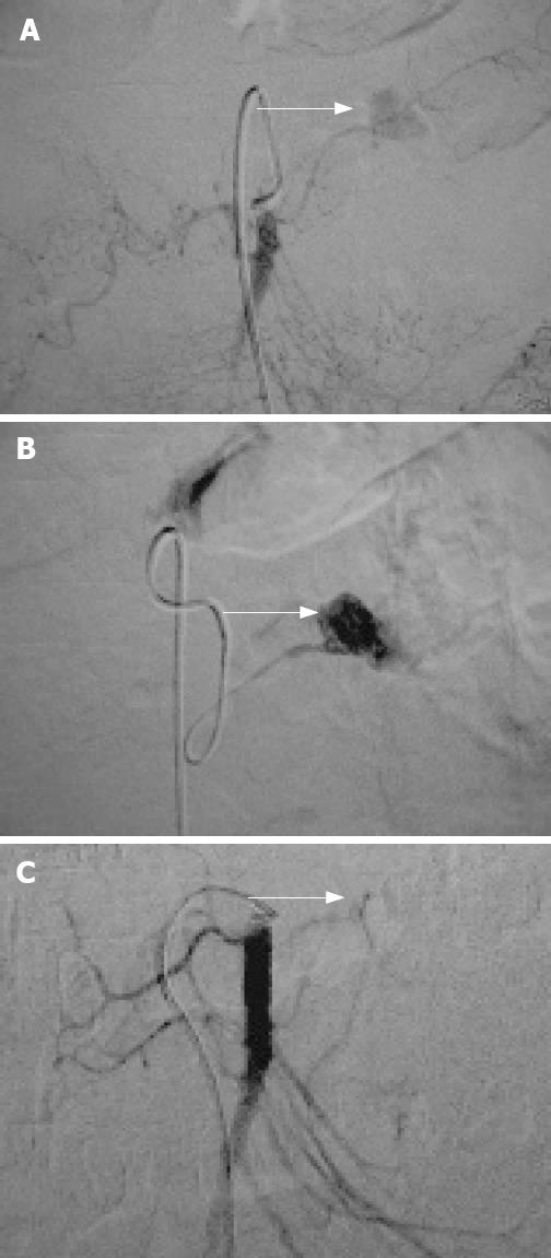
Figure 2 Angiography.
A: SMA angiography shows a pseudoaneurysm in a branch of the SMA with extravasation of the contrast (arrow); B: Subselective angiography illustrates extravasation of the contrast more clearly (arrow); C: After embolization of the damaged artery, there is cessation of extravasation of the contrast, with disappearance of the nidus (pseudoaneurysm) (arrow).
- Citation: He Q, Liu YQ, Liu Y, Guan YS. Acute necrotizing pancreatitis complicated with pancreatic pseudoaneurysm of the superior mesenteric artery: A case report. World J Gastroenterol 2008; 14(16): 2612-2614
- URL: https://www.wjgnet.com/1007-9327/full/v14/i16/2612.htm
- DOI: https://dx.doi.org/10.3748/wjg.14.2612


