Copyright
©2007 Baishideng Publishing Group Co.
World J Gastroenterol. Feb 28, 2007; 13(8): 1303-1305
Published online Feb 28, 2007. doi: 10.3748/wjg.v13.i8.1303
Published online Feb 28, 2007. doi: 10.3748/wjg.v13.i8.1303
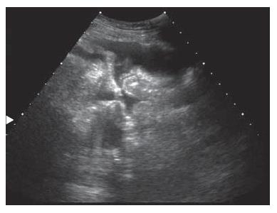
Figure 1 Ultrasonography of the abdomen revealed a moderate amount of ascites.
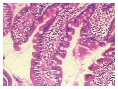
Figure 2 Histologic section of proximal duodenum showed inflammation with marked eosinophilic infiltration (HE, × 100).
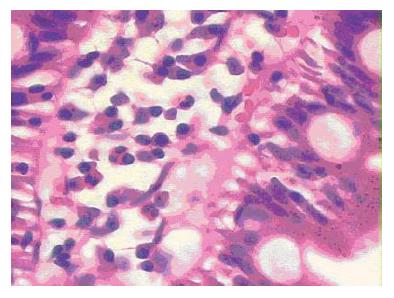
Figure 3 Histologic section of proximal duodenum showed inflammation with marked eosinophilic infiltration (HE, × 400).
- Citation: Zhou HB, Chen JM, Du Q. Eosinophilic gastroenteritis with ascites and hepatic dysfunction. World J Gastroenterol 2007; 13(8): 1303-1305
- URL: https://www.wjgnet.com/1007-9327/full/v13/i8/1303.htm
- DOI: https://dx.doi.org/10.3748/wjg.v13.i8.1303


