Copyright
©2007 Baishideng Publishing Group Co.
World J Gastroenterol. Feb 21, 2007; 13(7): 1119-1122
Published online Feb 21, 2007. doi: 10.3748/wjg.v13.i7.1119
Published online Feb 21, 2007. doi: 10.3748/wjg.v13.i7.1119
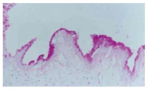
Figure 1 Specimens of chronic cholecystitis.
Many positive PAS materials appear in epithelial cells of gallbladder mucosa (PAS × 200).
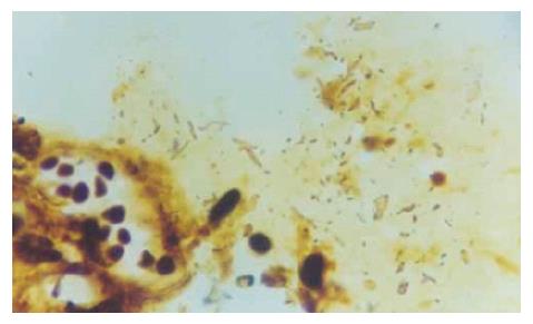
Figure 2 Helicobacter-like bacteria and inflammatory cells in mucus on gallbladder mucosa (WS × 200).
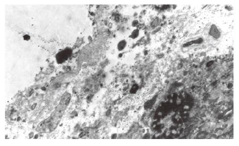
Figure 3 Electron microscopic images of H pylori on the epithelial cells of gallbladder (× 6000).
-
Citation: Chen DF, Hu L, Yi P, Liu WW, Fang DC, Cao H.
H pylori are associated with chronic cholecystitis. World J Gastroenterol 2007; 13(7): 1119-1122 - URL: https://www.wjgnet.com/1007-9327/full/v13/i7/1119.htm
- DOI: https://dx.doi.org/10.3748/wjg.v13.i7.1119


