Copyright
©2007 Baishideng Publishing Group Co.
World J Gastroenterol. Feb 14, 2007; 13(6): 970-972
Published online Feb 14, 2007. doi: 10.3748/wjg.v13.i6.970
Published online Feb 14, 2007. doi: 10.3748/wjg.v13.i6.970
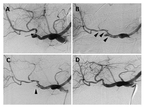
Figure 1 A: A severe stricture of a narrow segment in the proper hepatic artery and a bulging at the stump of the gastroduodenal artery were seen.
An arrow indicates a stricture; B: The stent-graft for an angioplasty was introduced through the stenotic segment to secure the hepatopetal arterial flow. Arrow heads indicate the stent-graft placed in the hepatic artery; C: After the stenting, embolization of the pseudoaneurysm was attempted to stop the inflow into the pseudoaneurysmal lumen using microcoils. An arrow head indicates microcoils placed in the pseudoaneurysmal lumen; D: The patency of the hepatic artery and disappearance of the pseudoaneurysm were confirmed by arteriography 8 d after the treatment.
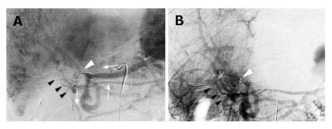
Figure 2 A: Portal phase of the celiac arteriography showed a severe stricture at the main trunk of the portal vein.
Hepatopetal collaterals through mesenteric veins were observed forming jejunal varices (black arrow heads) in the lifted jejunal limb. A white arrow head indicates the stricture and white arrows indicate the direction of blood flow; B: Portal venous phase of the superior mesenteric arteriogram also depicts hepatopetal collaterals and jejunal varices (black arrows) around the hepaticojejunostomy. White arrow heads indicate the stricture of the portal vein adjacent to the microcoils placed in the pseudoaneurysm.
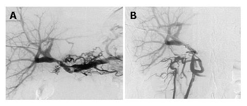
Figure 3 Portography after urokinase infusion showed a good portal blood flow and collaterals forming jejunal varices had disappeared.
A: Catheter tip was placed in the splenic vein; B: Catheter tip was placed in the superior mesenteric vein.
- Citation: Ichihara T, Sato T, Miyazawa H, Shibata S, Hashimoto M, Ishiyama K, Yamamoto Y. Stent placement is effective on both postoperative hepatic arterial pseudoaneurysm and subsequent portal vein stricture: A case report. World J Gastroenterol 2007; 13(6): 970-972
- URL: https://www.wjgnet.com/1007-9327/full/v13/i6/970.htm
- DOI: https://dx.doi.org/10.3748/wjg.v13.i6.970


