Copyright
©2007 Baishideng Publishing Group Co.
World J Gastroenterol. Feb 14, 2007; 13(6): 970-972
Published online Feb 14, 2007. doi: 10.3748/wjg.v13.i6.970
Published online Feb 14, 2007. doi: 10.3748/wjg.v13.i6.970
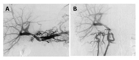
Figure 3 Portography after urokinase infusion showed a good portal blood flow and collaterals forming jejunal varices had disappeared.
A: Catheter tip was placed in the splenic vein; B: Catheter tip was placed in the superior mesenteric vein.
- Citation: Ichihara T, Sato T, Miyazawa H, Shibata S, Hashimoto M, Ishiyama K, Yamamoto Y. Stent placement is effective on both postoperative hepatic arterial pseudoaneurysm and subsequent portal vein stricture: A case report. World J Gastroenterol 2007; 13(6): 970-972
- URL: https://www.wjgnet.com/1007-9327/full/v13/i6/970.htm
- DOI: https://dx.doi.org/10.3748/wjg.v13.i6.970


