Copyright
©2007 Baishideng Publishing Group Co.
World J Gastroenterol. Feb 14, 2007; 13(6): 858-865
Published online Feb 14, 2007. doi: 10.3748/wjg.v13.i6.858
Published online Feb 14, 2007. doi: 10.3748/wjg.v13.i6.858
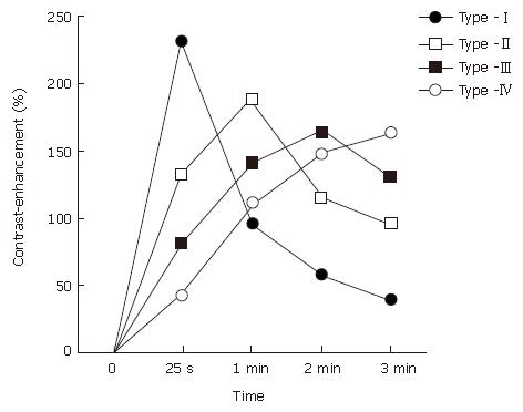
Figure 1 Patterns of the time-signal intensity curve (TIC) from dynamic contrast-enhanced magnetic resonance imaging of the pancreas.
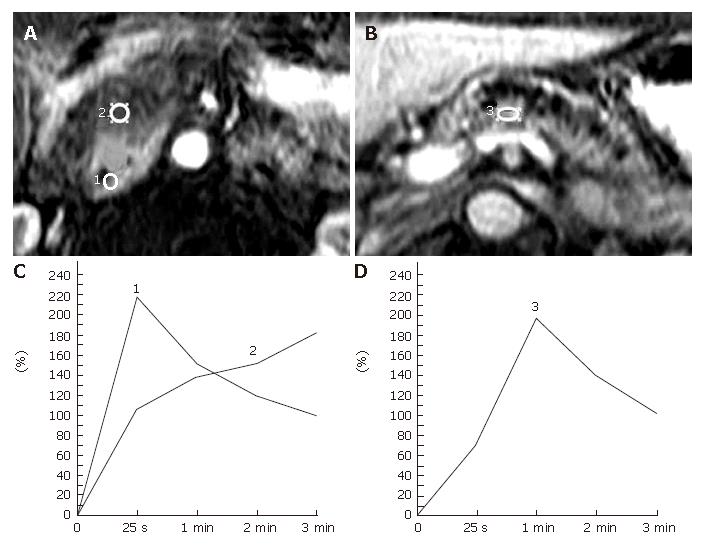
Figure 2 Representative pancreatic TIC profiles in patients with pancreatic ductal carcinoma developed in a normal pancreas.
A, B: Dynamic contrast-enhanced MRI images of the pancreas in a 59-year-old man with carcinoma of the head of the pancreas. The ROIs are placed at the pancreatic mass (No.2 ROI) and the non-tumorous pancreatic parenchyma both proximal (No.1 ROI) and distal (No.3 ROI) to the mass lesion; C: Pancreatic TICs obtained from the no.1 and no. 2 ROIs as in (A) demonstrate type-I and type-IV, respectively; D: Pancreatic TIC obtained from the No.3 ROI as in Figure 2B shows type-II.
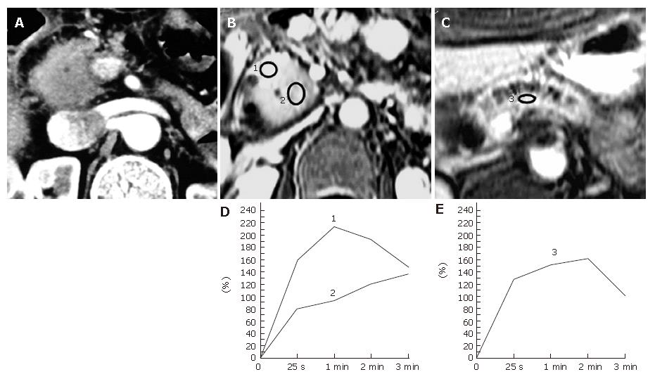
Figure 3 Pancreatic carcinoma occurring in a 67-year-old man with a longstanding chronic pancreatitis.
A: An abdominal contrast-enhanced CT image shows a focal enlargement of the head of the pancreas. The tumor-to-parenchymal attenuation difference is obscure. The patient underwent a laparotomy under a diagnosis of tumor-forming pancreatitis presenting with obstructive jaundice and was found to have pancreas head carcinoma during the operation; B, C: Dynamic contrast-enhanced MRI images of the pancreas. The ROIs are placed at the focally enlarged pancreas head (No.2 ROI), the proximal side of the head of the pancreas (No.1 ROI) , and the body of the pancreas (No.3 ROI); D: Pancreatic TICs obtained from the No.1 and No.2 ROIs as in (B) demonstrate type-II and type-IV, respectively; E: Pancreatic TIC obtained from the No.3 ROI as in (C) shows type-III.
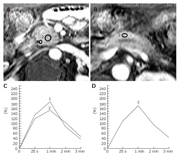
Figure 4 Tumor-forming pancreatitis in a 70-year-old man with a long history of alcohol abuse.
The patient underwent a pylorus-preserving pancreaticoduodenectomy together with lymphadenectomy for a suspected pancreas head carcinoma associated with obstructive jaundice and was confirmed to be chronic pancreatitis after surgery. A, B: Dynamic contrast-enhanced MRI images of the pancreas. The ROIs are placed at the pancreatic mass (No.2 ROI) and the pancreatic parenchyma both proximal (nNo.1 ROI) and distal (No.3 ROI) to the mass lesion; C: Both of the pancreatic TICs obtained from the No.1 and no.2 ROIs as in Figure 4A demonstrate type-II; D: Pancreatic TIC obtained from the No.3 ROI as in Figure 4B also shows type-II.
- Citation: Tajima Y, Kuroki T, Tsutsumi R, Isomoto I, Uetani M, Kanematsu T. Pancreatic carcinoma coexisting with chronic pancreatitis versus tumor-forming pancreatitis: Diagnostic utility of the time-signal intensity curve from dynamic contrast-enhanced MR imaging. World J Gastroenterol 2007; 13(6): 858-865
- URL: https://www.wjgnet.com/1007-9327/full/v13/i6/858.htm
- DOI: https://dx.doi.org/10.3748/wjg.v13.i6.858


