Copyright
©2007 Baishideng Publishing Group Co.
World J Gastroenterol. Dec 28, 2007; 13(48): 6603-6604
Published online Dec 28, 2007. doi: 10.3748/wjg.v13.i48.6603
Published online Dec 28, 2007. doi: 10.3748/wjg.v13.i48.6603
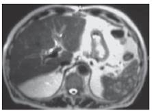
Figure 1 MRI: multiple hypointense lesions in the spleen (T2).
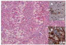
Figure 2 Vascular proliferation, well established (arrows), and very similar to normal spleen sinusoids.
A: Negative for CD8; B: Positive for histiomonocytic markers.
- Citation: Cosme &, Tejada &, Bujanda L, Vaquero M, Elorza JL, Ojeda E, Goikoetxea U. Littoral-cell angioma of the spleen: A case report. World J Gastroenterol 2007; 13(48): 6603-6604
- URL: https://www.wjgnet.com/1007-9327/full/v13/i48/6603.htm
- DOI: https://dx.doi.org/10.3748/wjg.v13.i48.6603


