Copyright
©2007 Baishideng Publishing Group Inc.
World J Gastroenterol. Dec 21, 2007; 13(47): 6436-6438
Published online Dec 21, 2007. doi: 10.3748/wjg.v13.i47.6436
Published online Dec 21, 2007. doi: 10.3748/wjg.v13.i47.6436
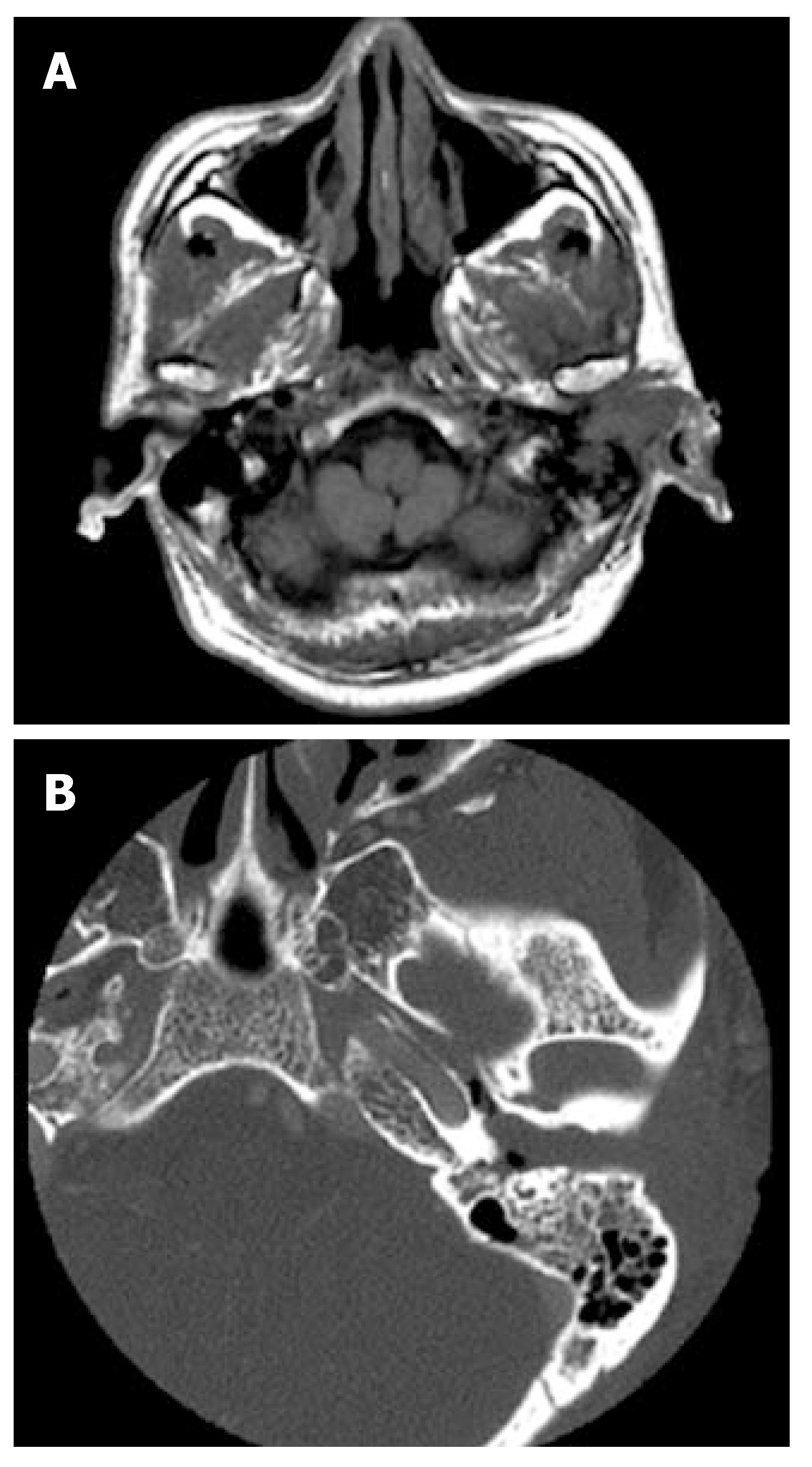
Figure 1 Axial T1-weighted magnetic resonance image of the head showing a huge mass in the left EAC with extension of the lesion into the mastoid (A), and axial CT of the temporal bone demonstrating the mass occupying the left EAC (B).
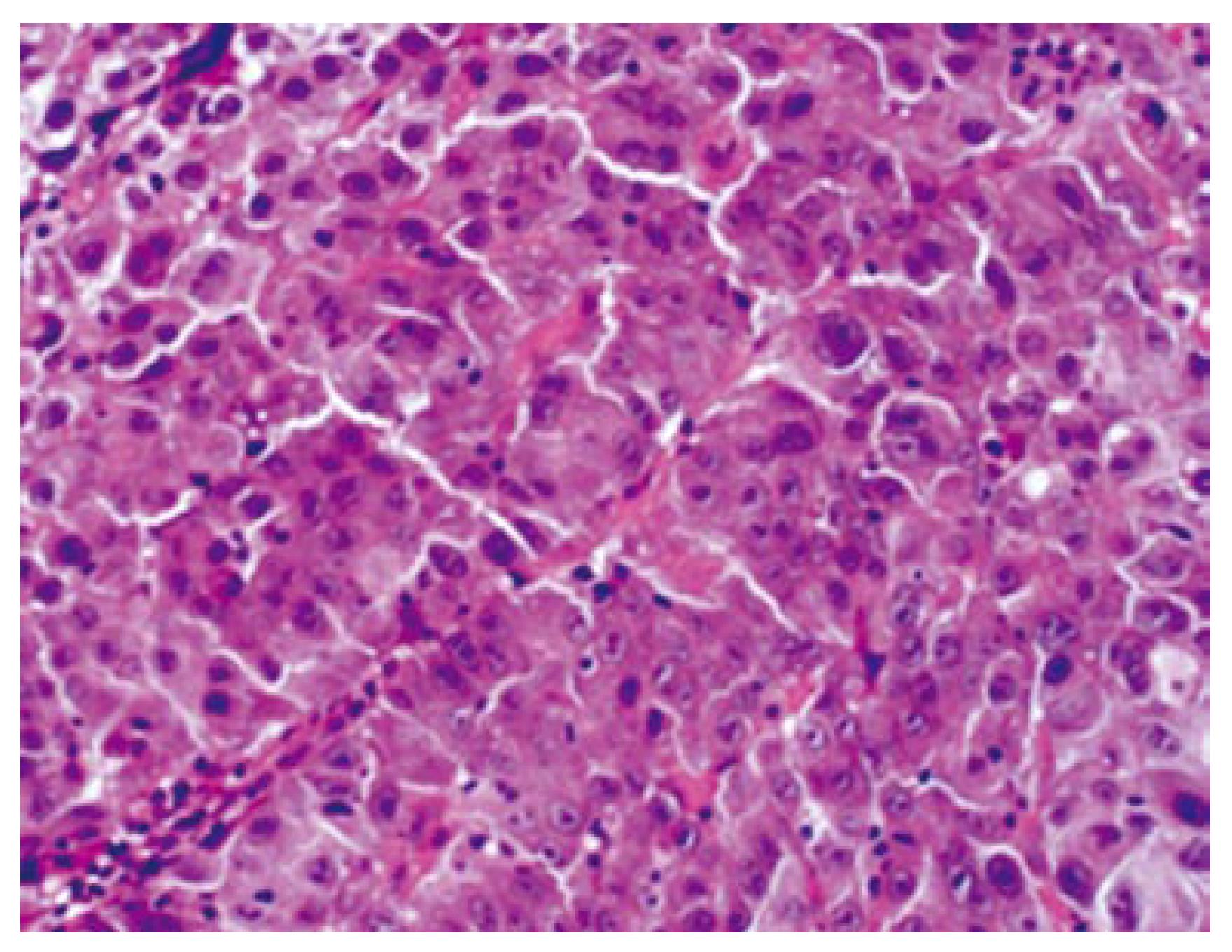
Figure 2 Microscopically, the lesion in the external auditory canal is composed of proliferating atypical epithelial cells forming a solid and thick trabecular arrangement with necrosis, and a fibrous stroma (hematoxylin/eosin stain, x 400).
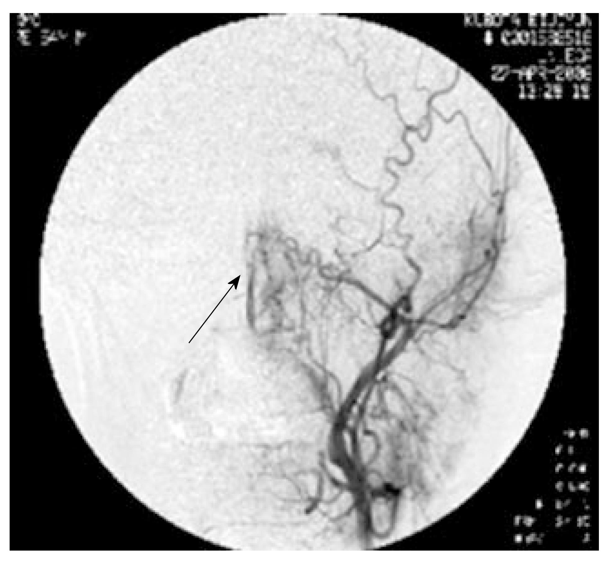
Figure 3 Angiography showing hypervascula-rity in the left external auditory canal supplied by the posterior auricular artery (the arrow indica-tes the tumor stain).
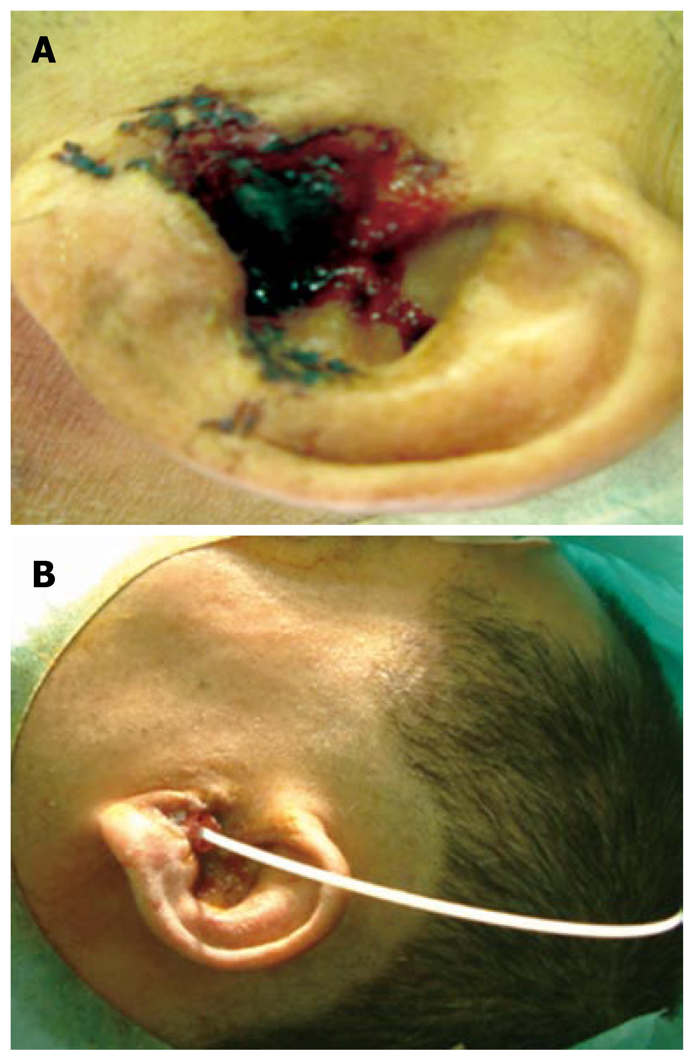
Figure 4 Metastatic hepatocellular carcinoma protruding from the left external auditory canal (A) and inserted and fixed catheter for the 192Ir remote afterloader system in the left external auditory canal after debulking surgery (B).
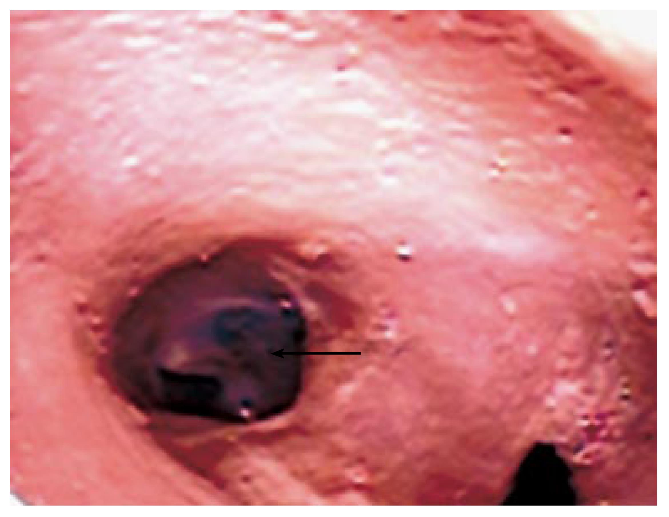
Figure 5 Fiberscopy of the external auditory canal 6 mo after treatment showing no evidence of residual tumor (the arrow indicates the left tympanic membrane).
- Citation: Yasumatsu R, Okura K, Sakiyama Y, Nakamuta M, Matsumura T, Uehara S, Yamamoto T, Komune S. Metastatic hepatocellular carcinoma of the external auditory canal. World J Gastroenterol 2007; 13(47): 6436-6438
- URL: https://www.wjgnet.com/1007-9327/full/v13/i47/6436.htm
- DOI: https://dx.doi.org/10.3748/wjg.v13.i47.6436


