Copyright
©2007 Baishideng Publishing Group Inc.
World J Gastroenterol. Dec 21, 2007; 13(47): 6436-6438
Published online Dec 21, 2007. doi: 10.3748/wjg.v13.i47.6436
Published online Dec 21, 2007. doi: 10.3748/wjg.v13.i47.6436
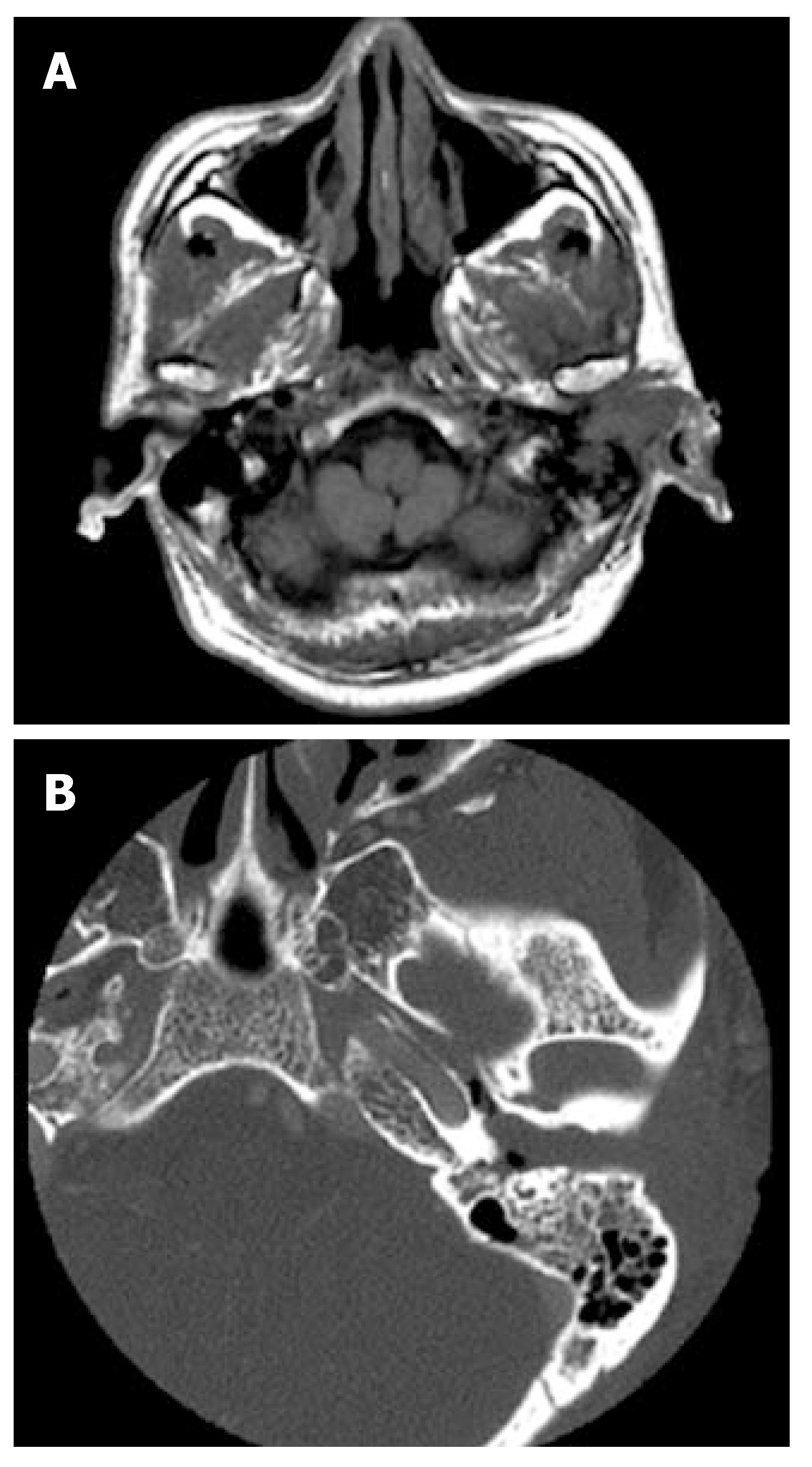
Figure 1 Axial T1-weighted magnetic resonance image of the head showing a huge mass in the left EAC with extension of the lesion into the mastoid (A), and axial CT of the temporal bone demonstrating the mass occupying the left EAC (B).
- Citation: Yasumatsu R, Okura K, Sakiyama Y, Nakamuta M, Matsumura T, Uehara S, Yamamoto T, Komune S. Metastatic hepatocellular carcinoma of the external auditory canal. World J Gastroenterol 2007; 13(47): 6436-6438
- URL: https://www.wjgnet.com/1007-9327/full/v13/i47/6436.htm
- DOI: https://dx.doi.org/10.3748/wjg.v13.i47.6436


