Copyright
©2007 Baishideng Publishing Group Co.
World J Gastroenterol. Nov 28, 2007; 13(44): 5954-5956
Published online Nov 28, 2007. doi: 10.3748/wjg.v13.i44.5954
Published online Nov 28, 2007. doi: 10.3748/wjg.v13.i44.5954
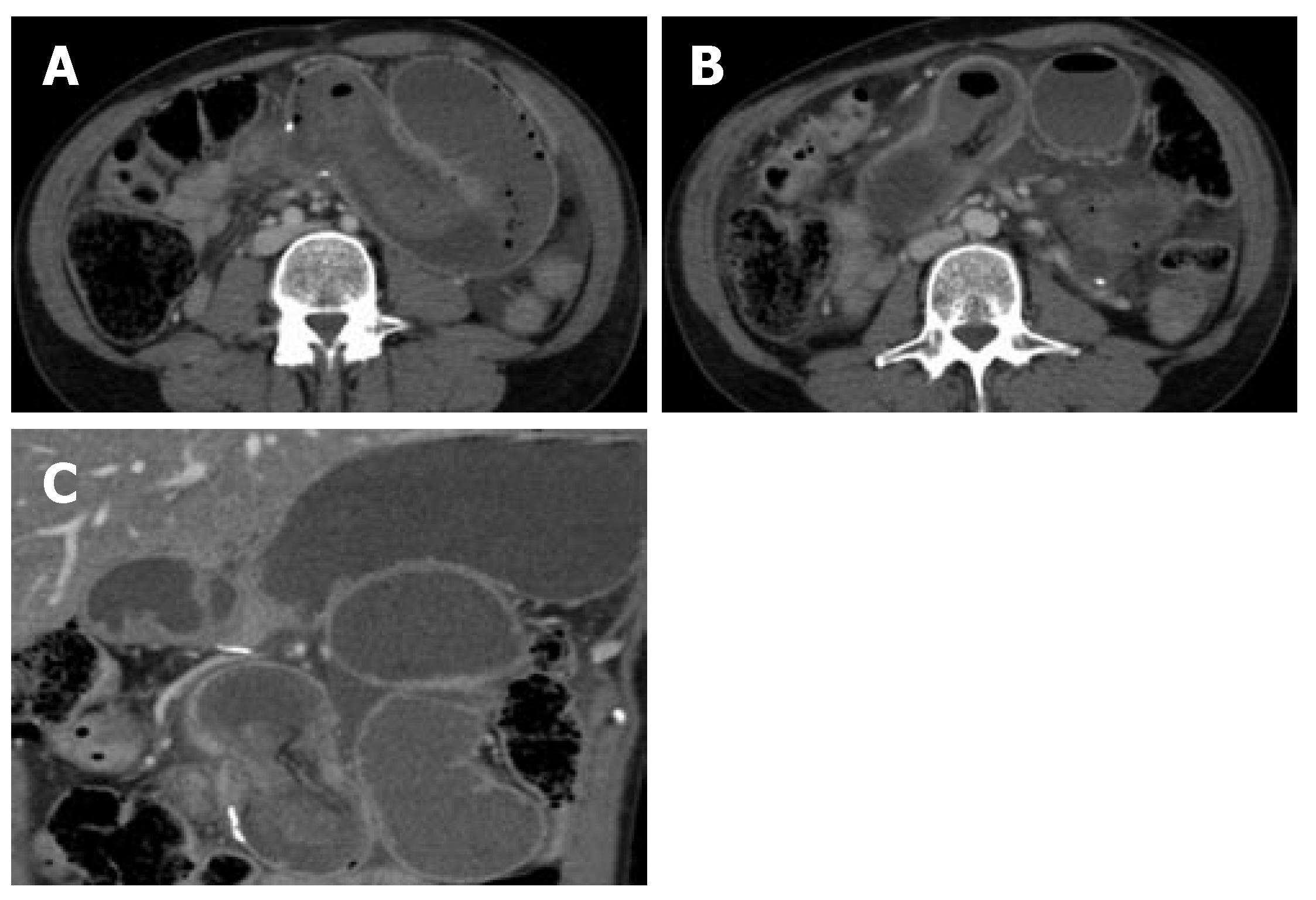
Figure 1 Axial (A and B) and coronal (C) CT images of the abdomen following intravenous contrast administration, which show large dilated loops of small bowel proximal to the intussusception.
The intussuscepted bowel entered the more distal jejunum via the jejunal anastomotic site, which is evident due to the presence of surgical clips.
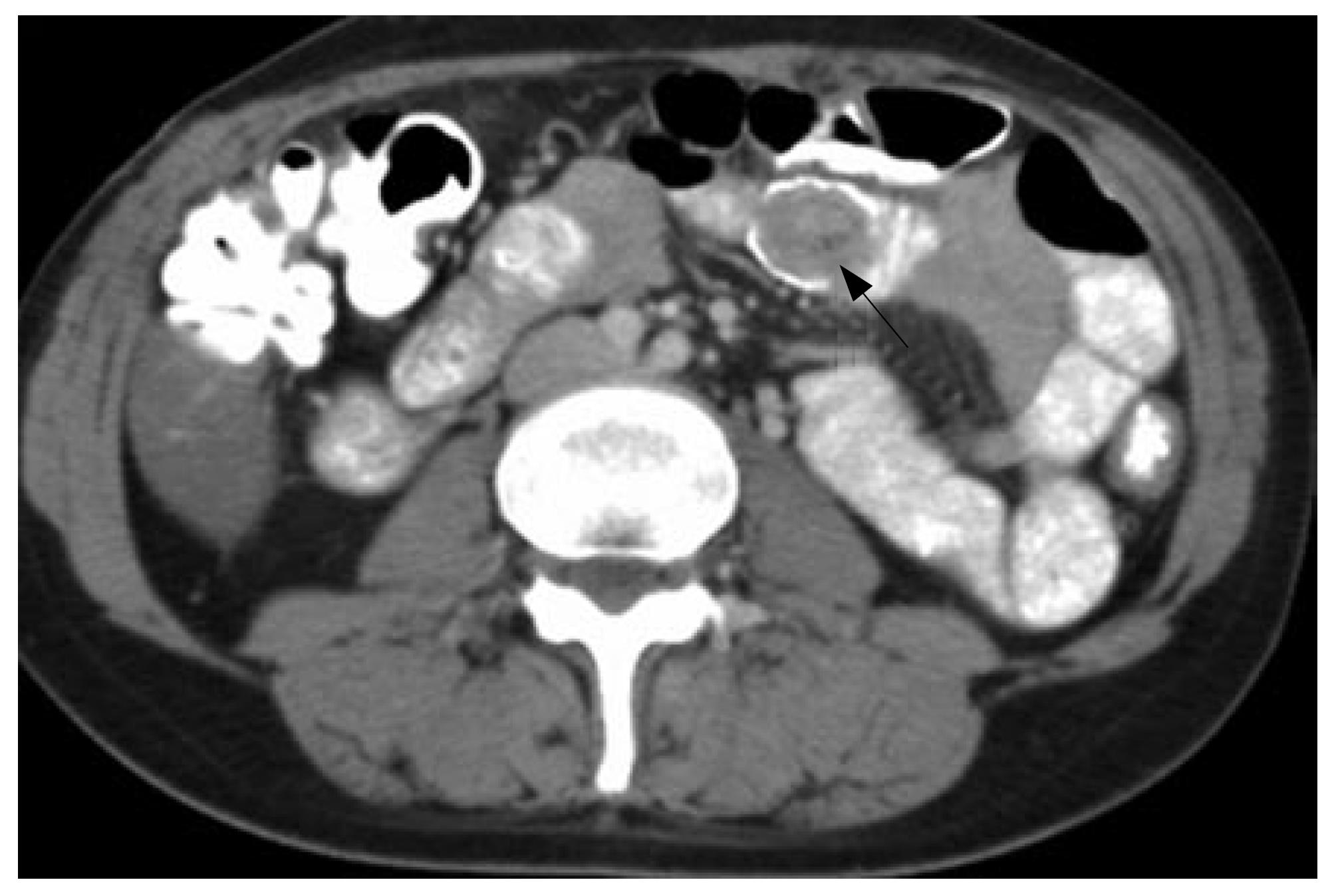
Figure 2 Abdominal CT scan from prior hospital visits, which reveals milder bowel intussusception prior to the patient’s last admission.
- Citation: Gigena M, Villar HV, Knowles NG, Cunningham JT, Outwater EK, Leon Jr LR. Antegrade bowel intussusception after remote Whipple and Puestow procedures for treatment of pancreas divisum. World J Gastroenterol 2007; 13(44): 5954-5956
- URL: https://www.wjgnet.com/1007-9327/full/v13/i44/5954.htm
- DOI: https://dx.doi.org/10.3748/wjg.v13.i44.5954


