Copyright
©2007 Baishideng Publishing Group Inc.
World J Gastroenterol. Nov 21, 2007; 13(43): 5783-5786
Published online Nov 21, 2007. doi: 10.3748/wjg.v13.i43.5783
Published online Nov 21, 2007. doi: 10.3748/wjg.v13.i43.5783
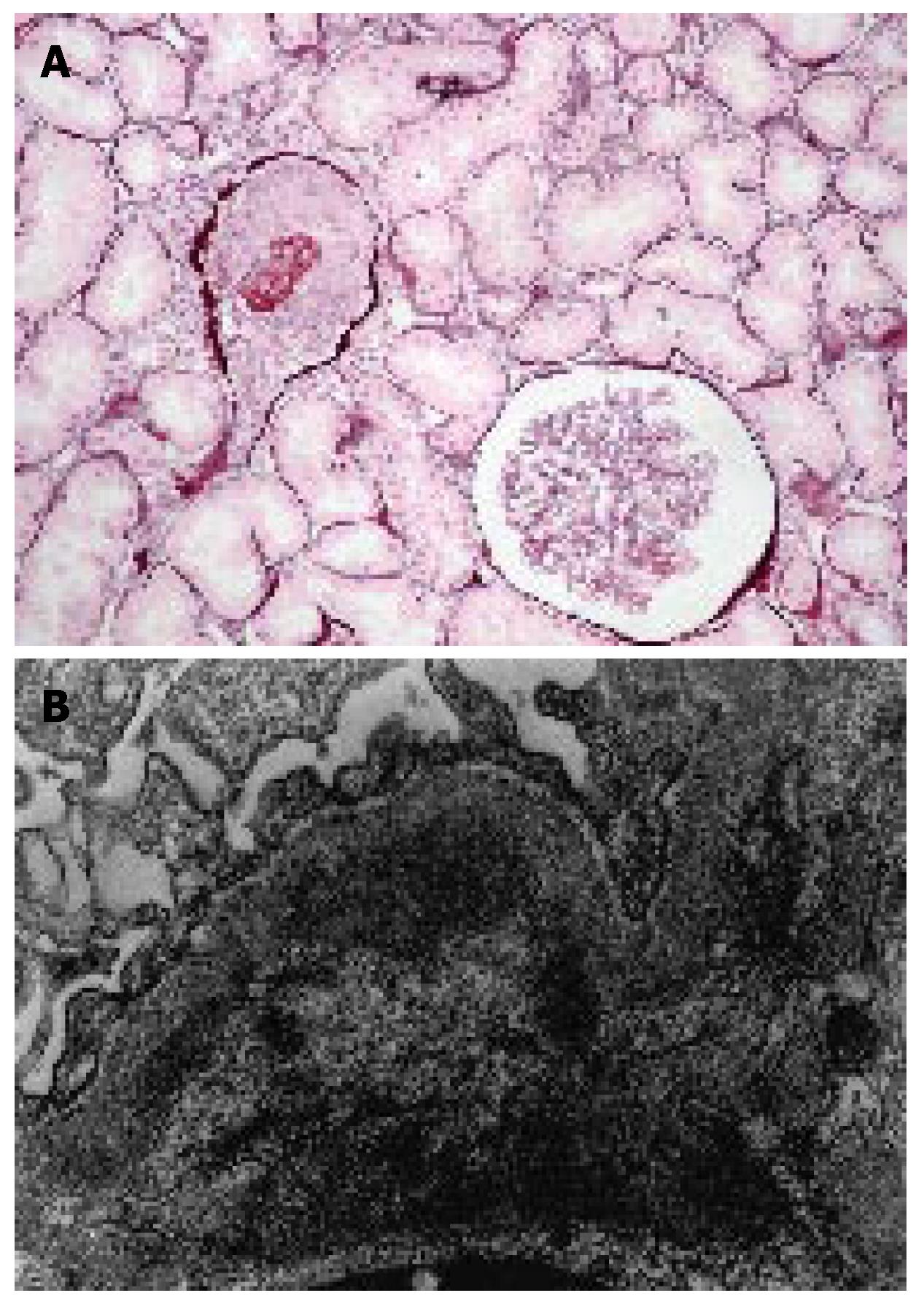
Figure 1 A: Light microscopy study showing endocapillary and extracapillary proliferation.
A large cellular crescend is observed in the left upper glomerulus (silver methoxamine stain, × 200); B: Electron-microscopic study showing intramembranous and paramesangial electron-dense deposits (× 13000).
- Citation: Kalambokis G, Christou L, Stefanou D, Arkoumani E, Tsianos EV. Association of liver cirrhosis related IgA nephropathy with portal hypertension. World J Gastroenterol 2007; 13(43): 5783-5786
- URL: https://www.wjgnet.com/1007-9327/full/v13/i43/5783.htm
- DOI: https://dx.doi.org/10.3748/wjg.v13.i43.5783


