Copyright
©2007 Baishideng Publishing Group Inc.
World J Gastroenterol. Nov 21, 2007; 13(43): 5718-5724
Published online Nov 21, 2007. doi: 10.3748/wjg.v13.i43.5718
Published online Nov 21, 2007. doi: 10.3748/wjg.v13.i43.5718
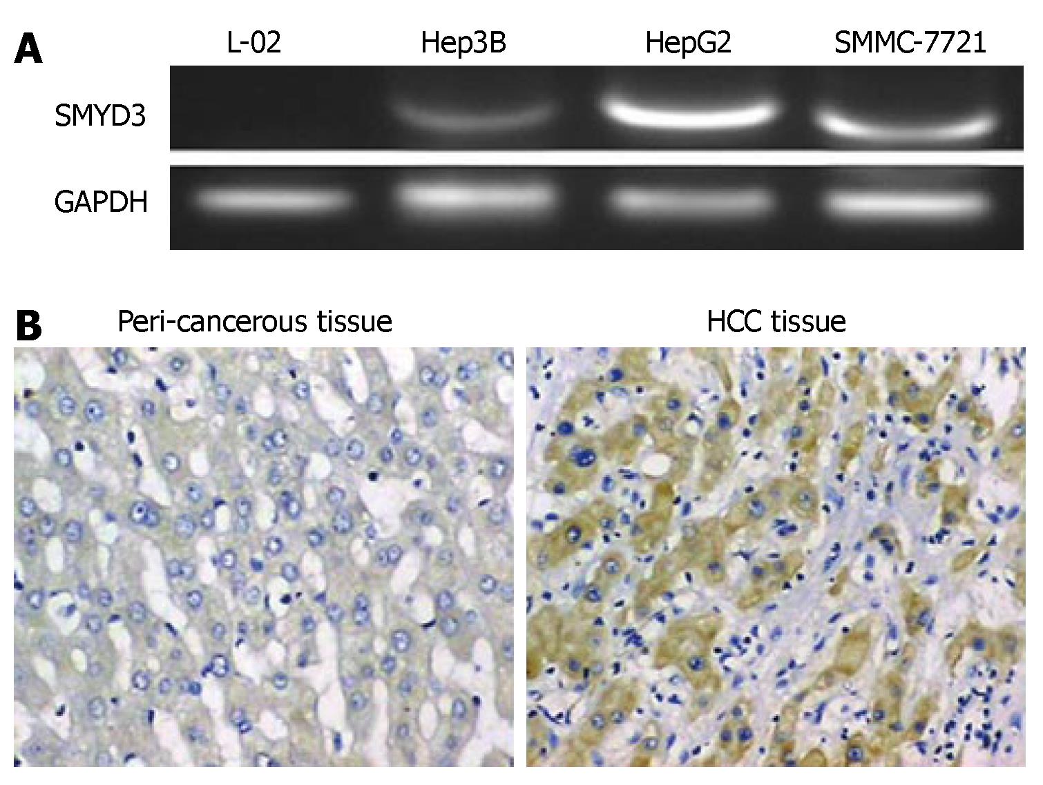
Figure 1 Enhanced expression of SMYD3 in HCC.
A: The expression of SMYD3 genes was determined by RT-PCR and GAPDH served as an internal control; B: Representative images of immunohistochemical staining of accumulated SMYD3 protein in HCC tissue, but not in peri-cancerous tissue.
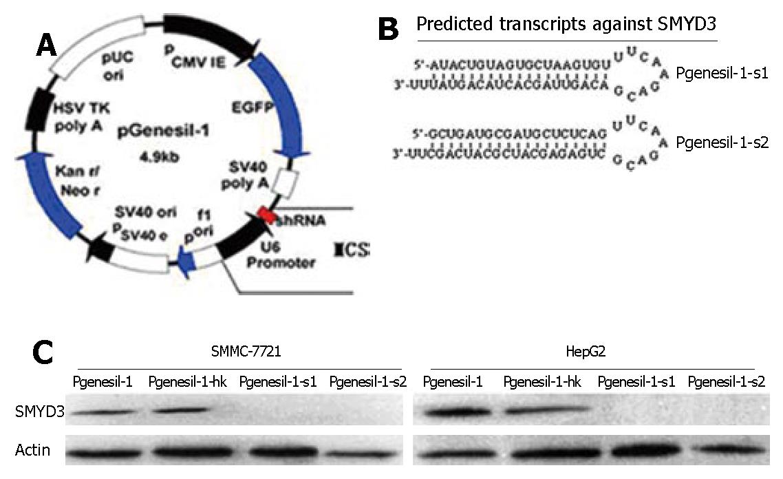
Figure 2 RNAi specifically inhibits SMYD3 expression in HCC cell lines.
A: Schematic drawing of the Pgenesil-1 vector; B: The predicted secondary structures of the Pgenesil-1-s1 and Pgenesil-1-s2 transcripts target SMYD3 are shown; C: 48h after transfection, Western blot analysis showed the inhibitory effect of plasmids expressing SMYD3 shRNAs in HepG2 and SMMC-7721 cells.
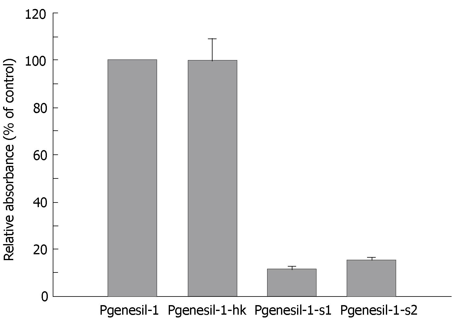
Figure 3 Inhibition of SMYD3 reduces hepatoma cell proliferation (MTT assays).
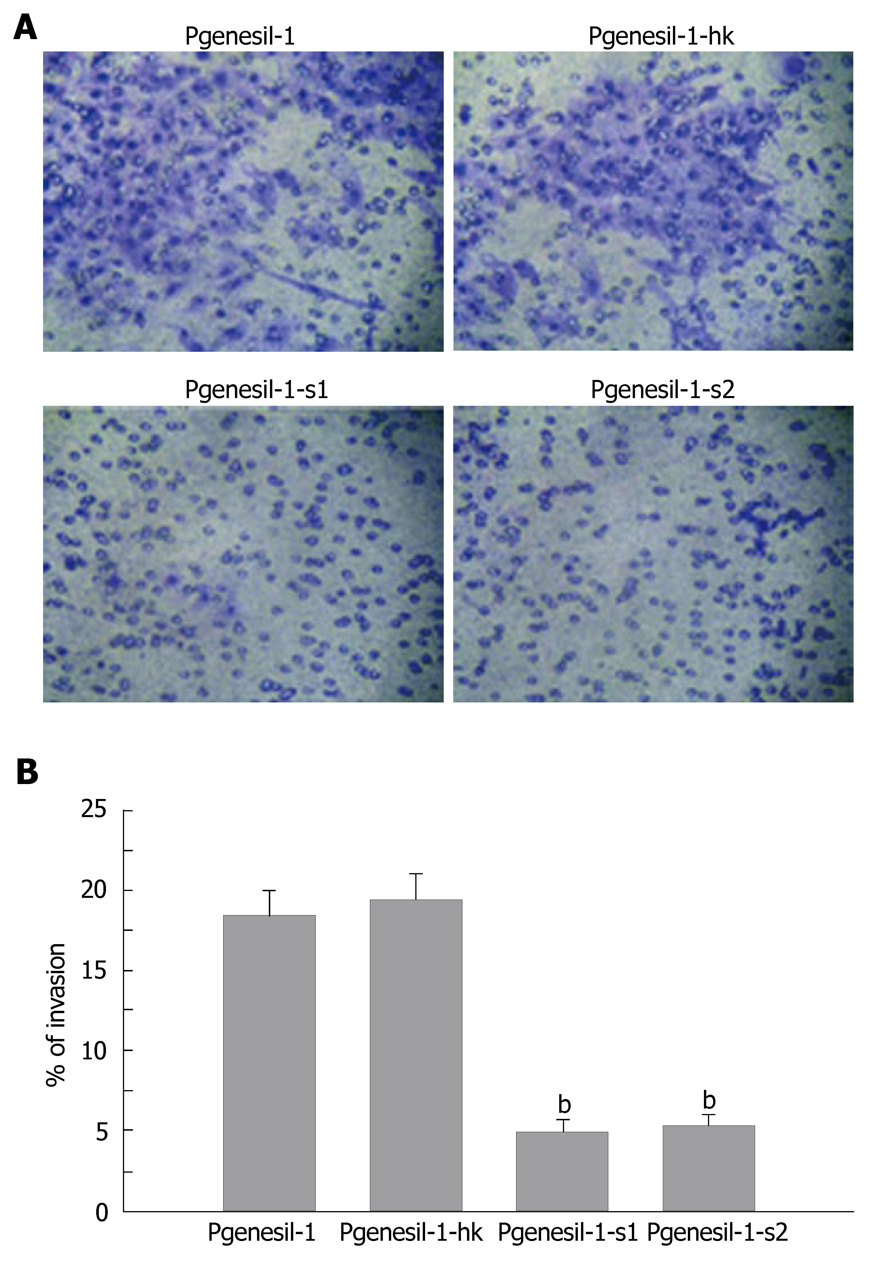
Figure 4 Inhibition of SMYD3 reduces hepatoma cell migration.
A: Representative cells (blue) invade through Matrigel and membrane; B: The percentage of cells successfully passing through the matrigel and membrane (P < 0.01).
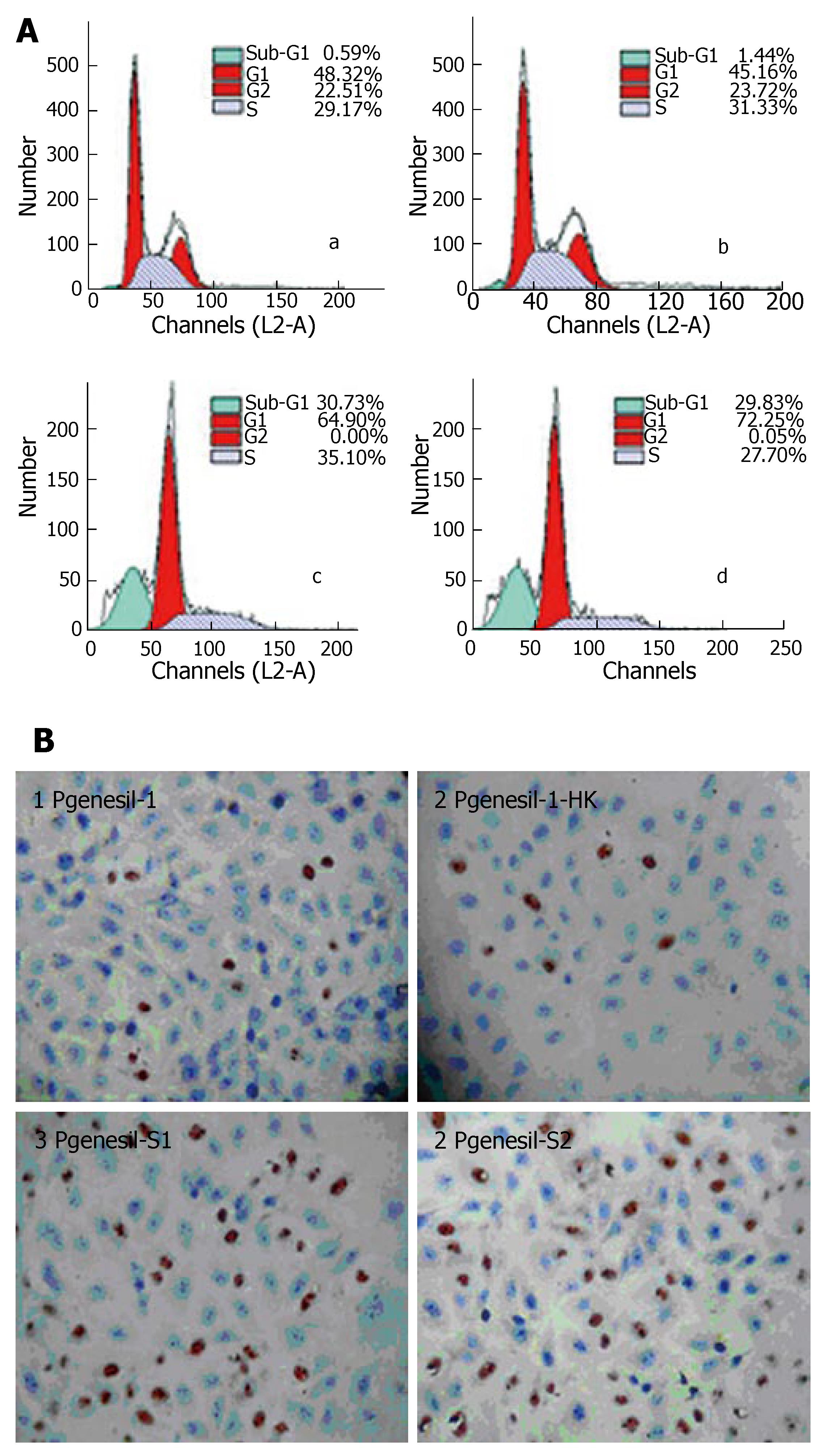
Figure 5 Inhibition of SMYD3 promotes apoptosis in hepatoma cells upon serum deprivation.
A: FCM analysis of HepG2 cells 48 h after transfection (P < 0.01). B: TUNEL assay of cell apoptosis with siRNA suppression of SMYD3 expression in HepG2 (brown staining cells) (P < 0.01).
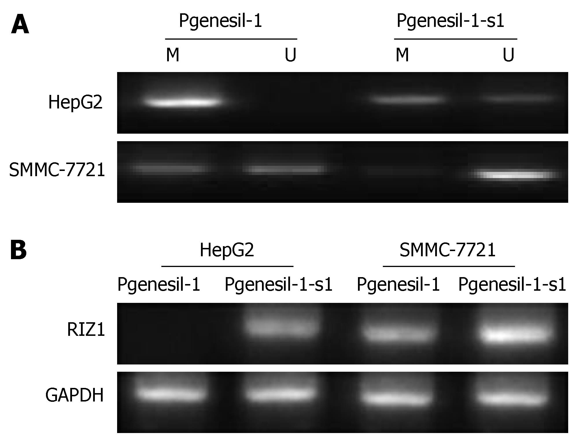
Figure 6 Inhibition of SMYD3 de-methylates RIZ1 promoter CpG islands and reexpresses RIZ1 in hepatoma Cells.
A: Methylation specific PCR (MSP) assay of RIZ1 promoter. M: Methylated; U: Unmethylated; B: RIZ1 mRNA expression, GAPDH served as internal control (RT-PCR).
- Citation: Chen LB, Xu JY, Yang Z, Wang GB. Silencing SMYD3 in hepatoma demethylates RIZI promoter induces apoptosis and inhibits cell proliferation and migration. World J Gastroenterol 2007; 13(43): 5718-5724
- URL: https://www.wjgnet.com/1007-9327/full/v13/i43/5718.htm
- DOI: https://dx.doi.org/10.3748/wjg.v13.i43.5718


