Copyright
©2007 Baishideng Publishing Group Inc.
World J Gastroenterol. Nov 7, 2007; 13(41): 5533-5536
Published online Nov 7, 2007. doi: 10.3748/wjg.v13.i41.5533
Published online Nov 7, 2007. doi: 10.3748/wjg.v13.i41.5533
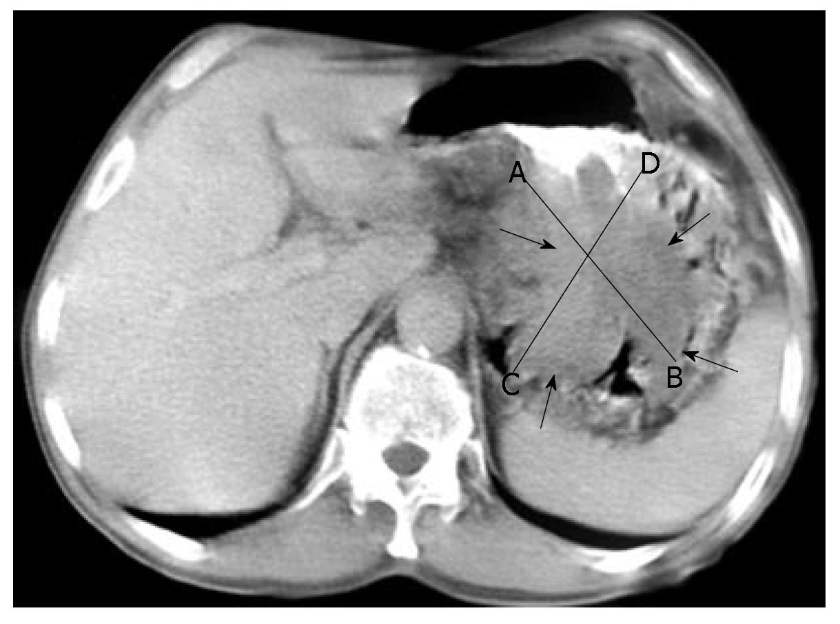
Figure 1 CT scan of the abdomen showing tumor (arrows).
AB, CD: tumor diameters.
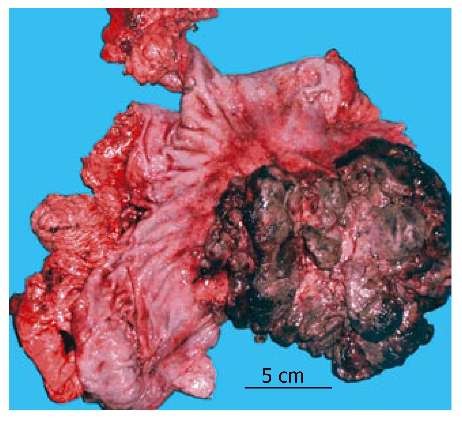
Figure 2 Macroscopic specimen obtained during surgery.
Tumor underwent central necrosis, and a hemorrhagic zone is visible on the periphery.
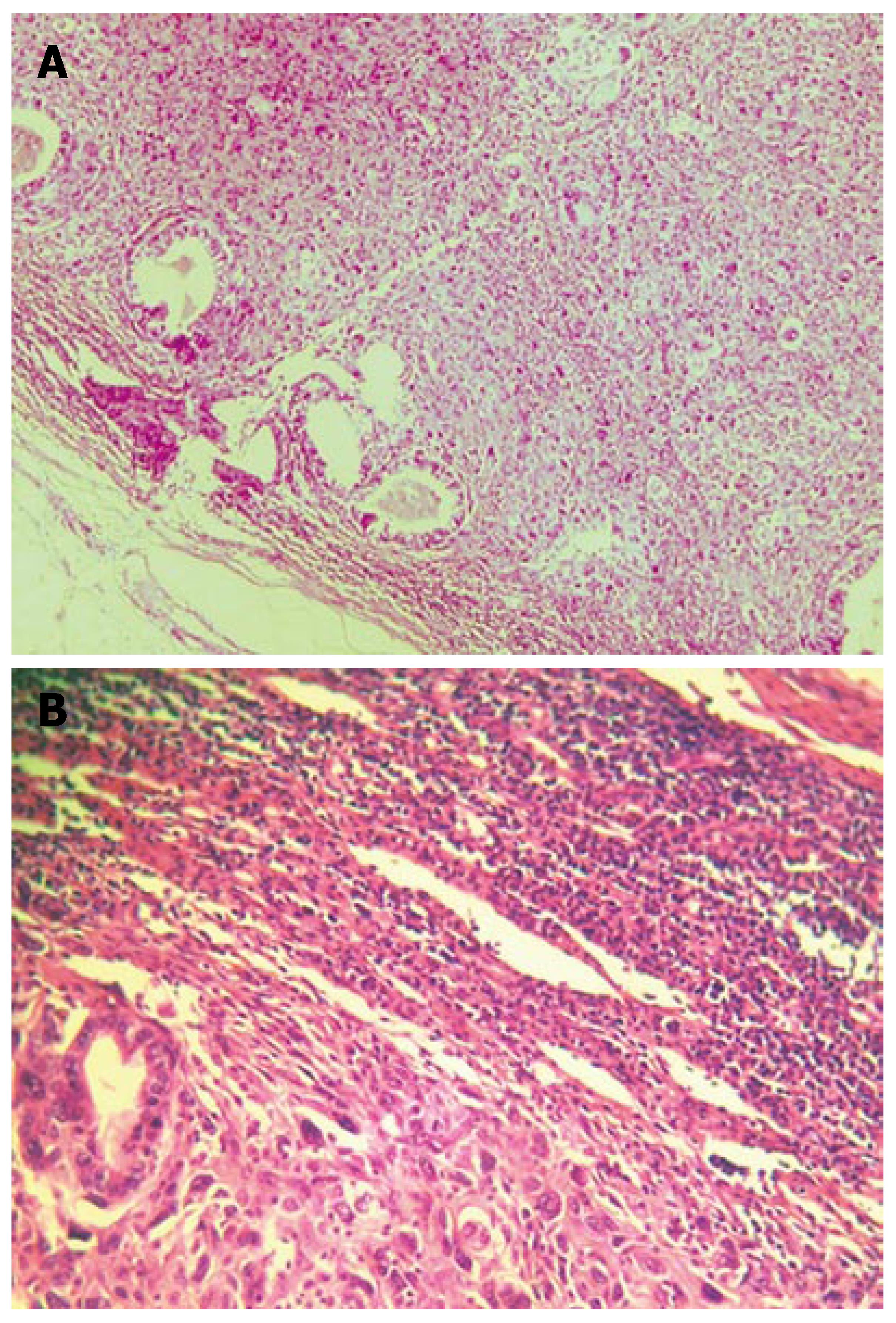
Figure 3 Massive lymph node infiltration with tumor cells.
Only a few tubules can be seen on the lymph-node periphery. In between and all around, large polygonal cells are haphazardly arranged (sarcomatous component). This appearance resembles that of the main tumor mass. A: hematoxylin-eosin (HE), × 50; B: HE, × 100.
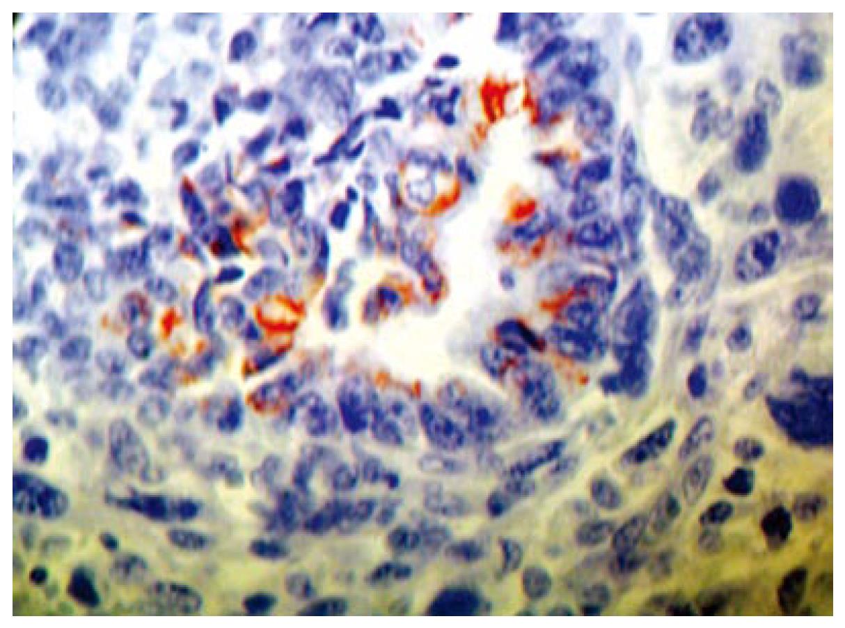
Figure 4 Cytokeratin-18-positive epithelial cells arranged in tubules (× 400).
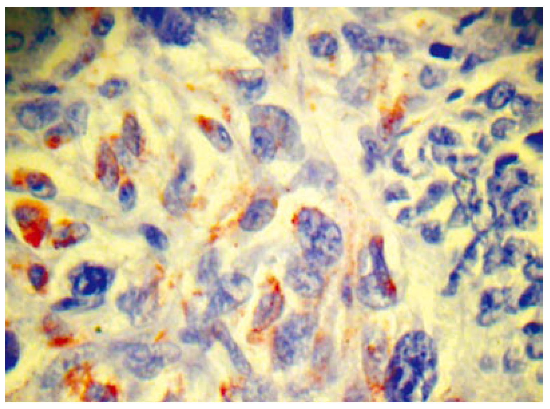
Figure 5 Vimentin-positive polygonal tumor cells (× 400).
- Citation: Randjelovic T, Filipovic B, Babic D, Cemerikic V, Filipovic B. Carcinosarcoma of the stomach: A case report and review of the literature. World J Gastroenterol 2007; 13(41): 5533-5536
- URL: https://www.wjgnet.com/1007-9327/full/v13/i41/5533.htm
- DOI: https://dx.doi.org/10.3748/wjg.v13.i41.5533


