Copyright
©2007 Baishideng Publishing Group Inc.
World J Gastroenterol. Nov 7, 2007; 13(41): 5525-5526
Published online Nov 7, 2007. doi: 10.3748/wjg.v13.i41.5525
Published online Nov 7, 2007. doi: 10.3748/wjg.v13.i41.5525
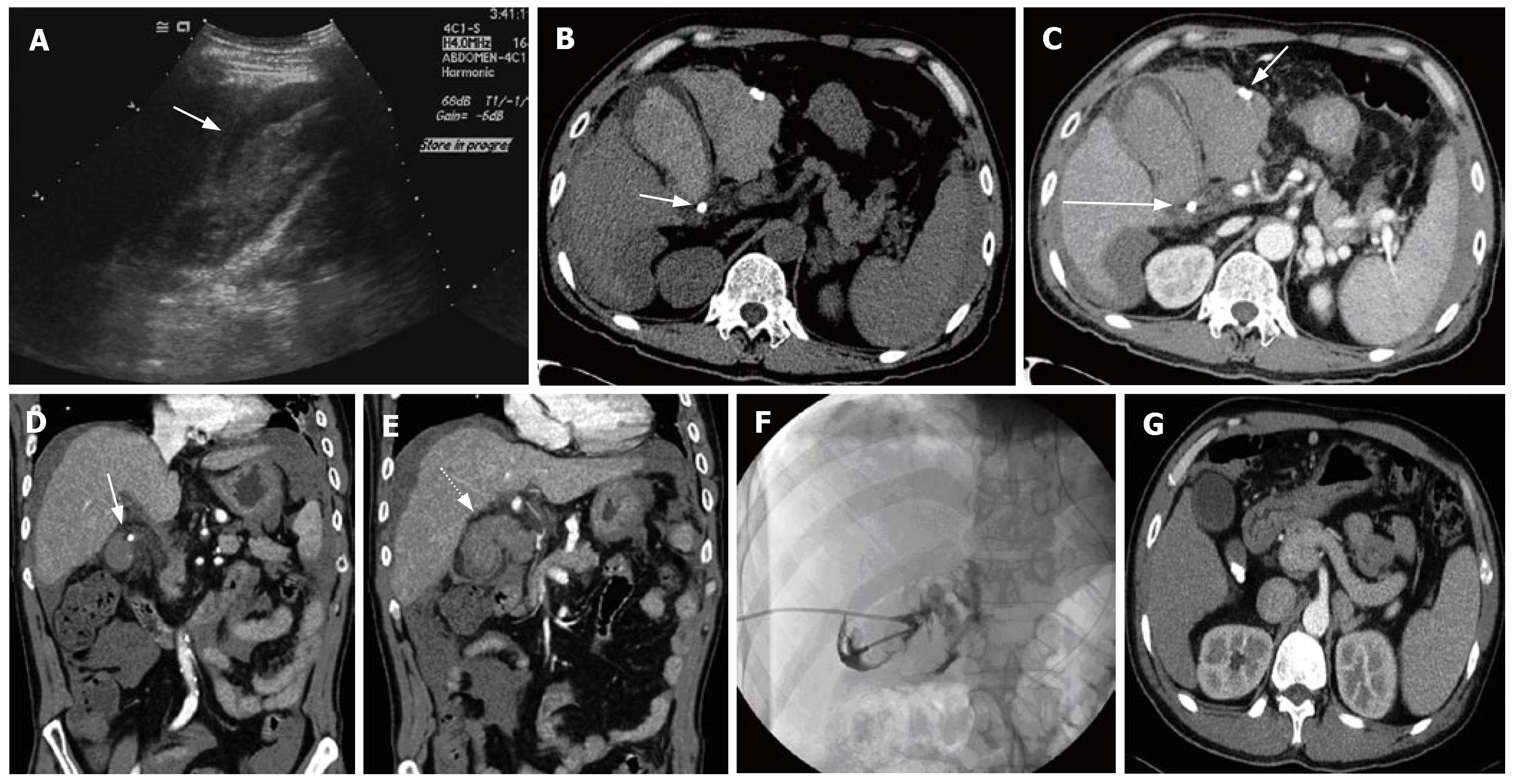
Figure 1 A 55-year-old man with right upper quadrant pain.
US images (A) demonstrate heterogeneous, highly echogenic material, both within and outside the gallbladder lumen (arrows), with a positive sonographic Murphy's sign. Non-contrast (B) and contrast-enhanced (C) transverse CT images show high-attenuation (46-61HU) material, both in the gallbladder lumen and pericholecystic space. One stone is seen in the cystic duct (long arrow) and calcified material (with the same appearance as the cystic duct stone) is seen in the fluid collected (short arrow) around the gallbladder. Contrast-enhanced coronal CT images (D, E) show well the impacted cystic-duct stone (arrow), and the mucosal defect with continuation of hemorrhage (dotted arrow). PTGBD (F) with cholecystography demonstrates contrast leakage from the gallbladder. Contrast-enhanced transverse CT images (G) taken 2 mo before the current attack show multiple stones in the gallbladder neck without complications.
- Citation: Kim YC, Park MS, Chung YE, Lim JS, Kim MJ, Kim KW. Gallstone spillage caused by spontaneously perforated hemorrhagic cholecystitis. World J Gastroenterol 2007; 13(41): 5525-5526
- URL: https://www.wjgnet.com/1007-9327/full/v13/i41/5525.htm
- DOI: https://dx.doi.org/10.3748/wjg.v13.i41.5525


