Copyright
©2007 Baishideng Publishing Group Inc.
World J Gastroenterol. Oct 28, 2007; 13(40): 5400-5402
Published online Oct 28, 2007. doi: 10.3748/wjg.v13.i40.5400
Published online Oct 28, 2007. doi: 10.3748/wjg.v13.i40.5400
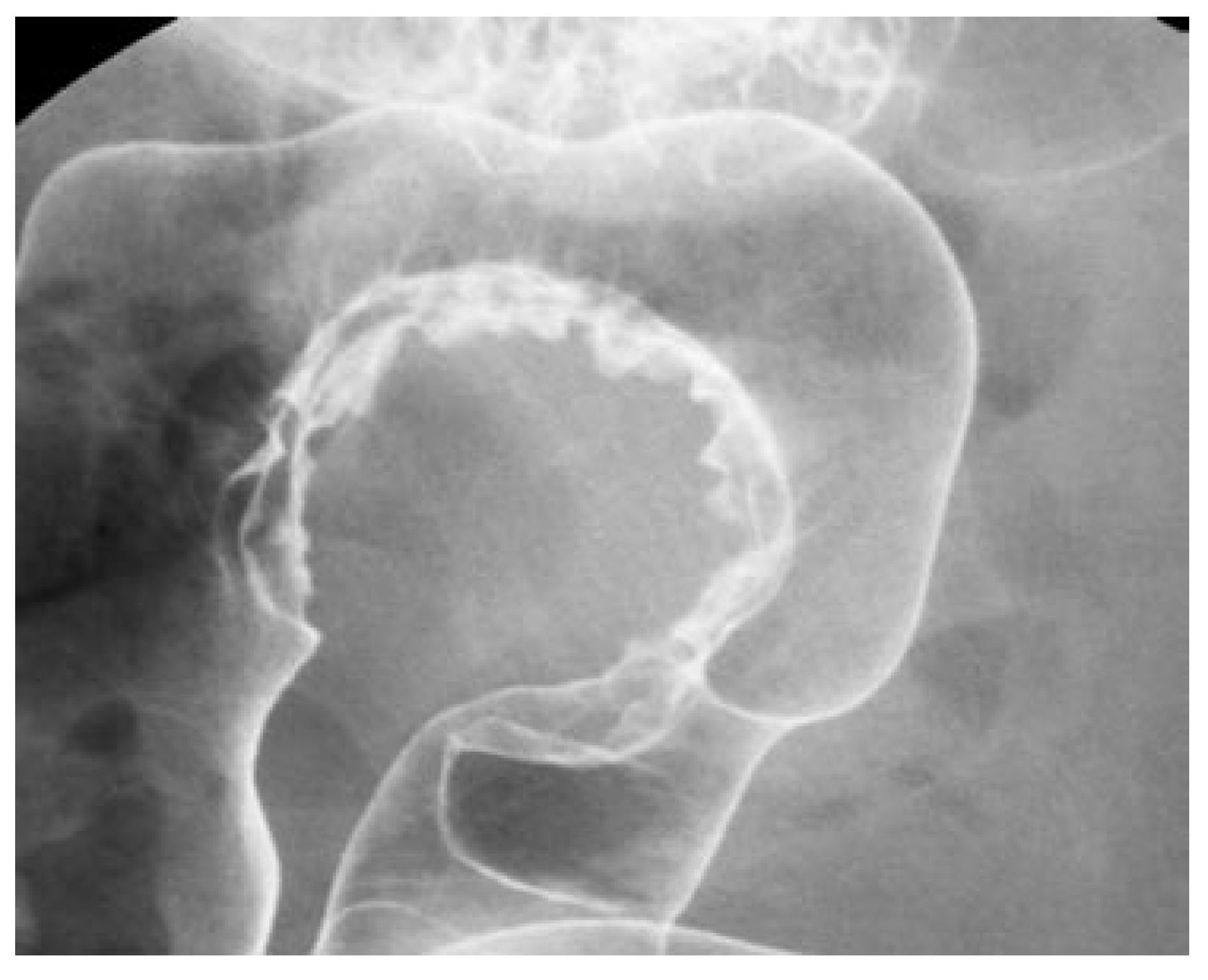
Figure 1 Barium enema shows sigmoidal stenosis with smooth inner lumen.
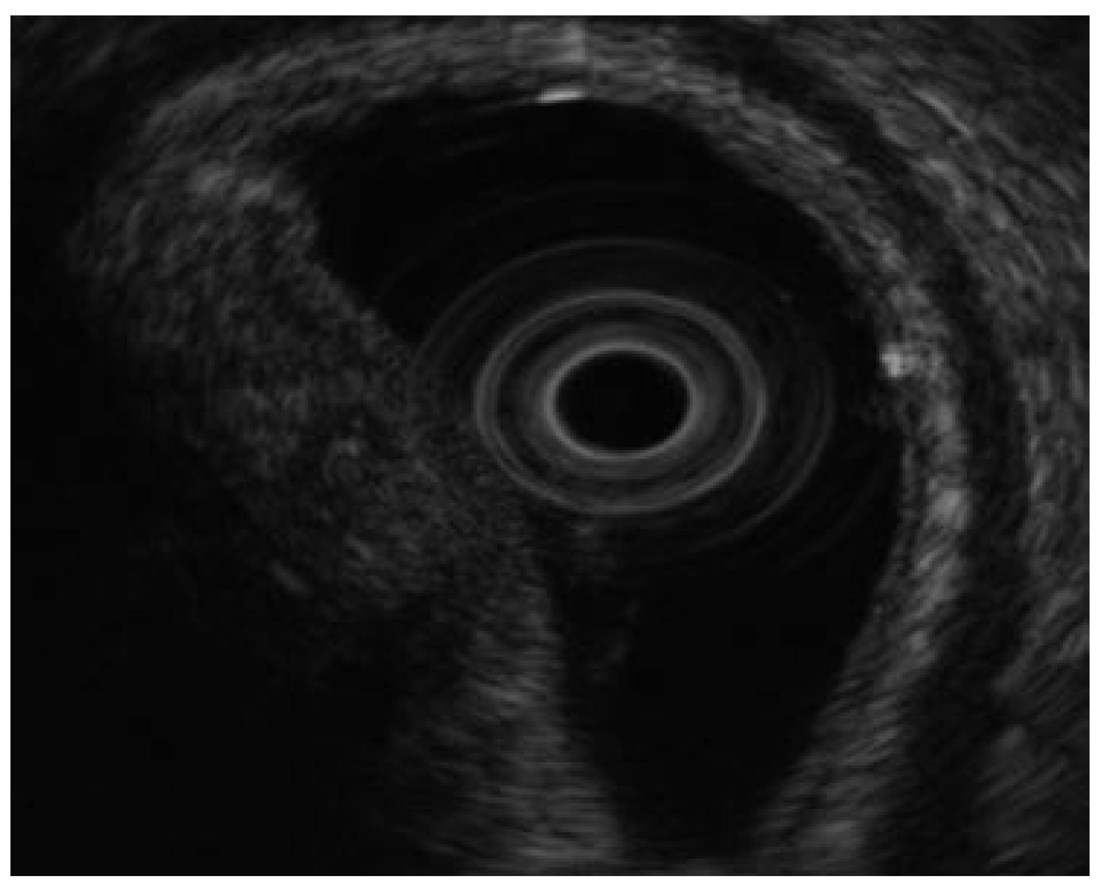
Figure 2 EUS shows iso to high echoic submucosal tumor in the 3rd echoic layer.
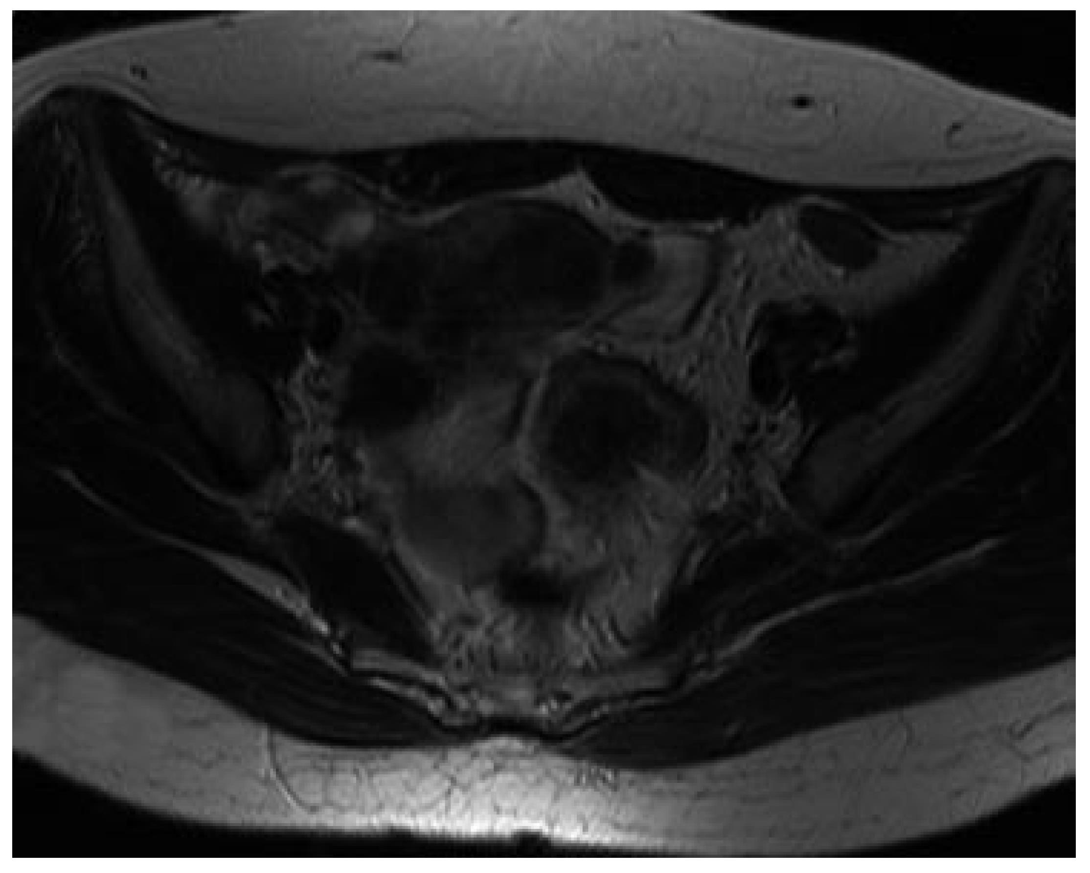
Figure 3 MRI (T2 weighted image) shows a sigmoidal tumor lesion with low signal intensity.
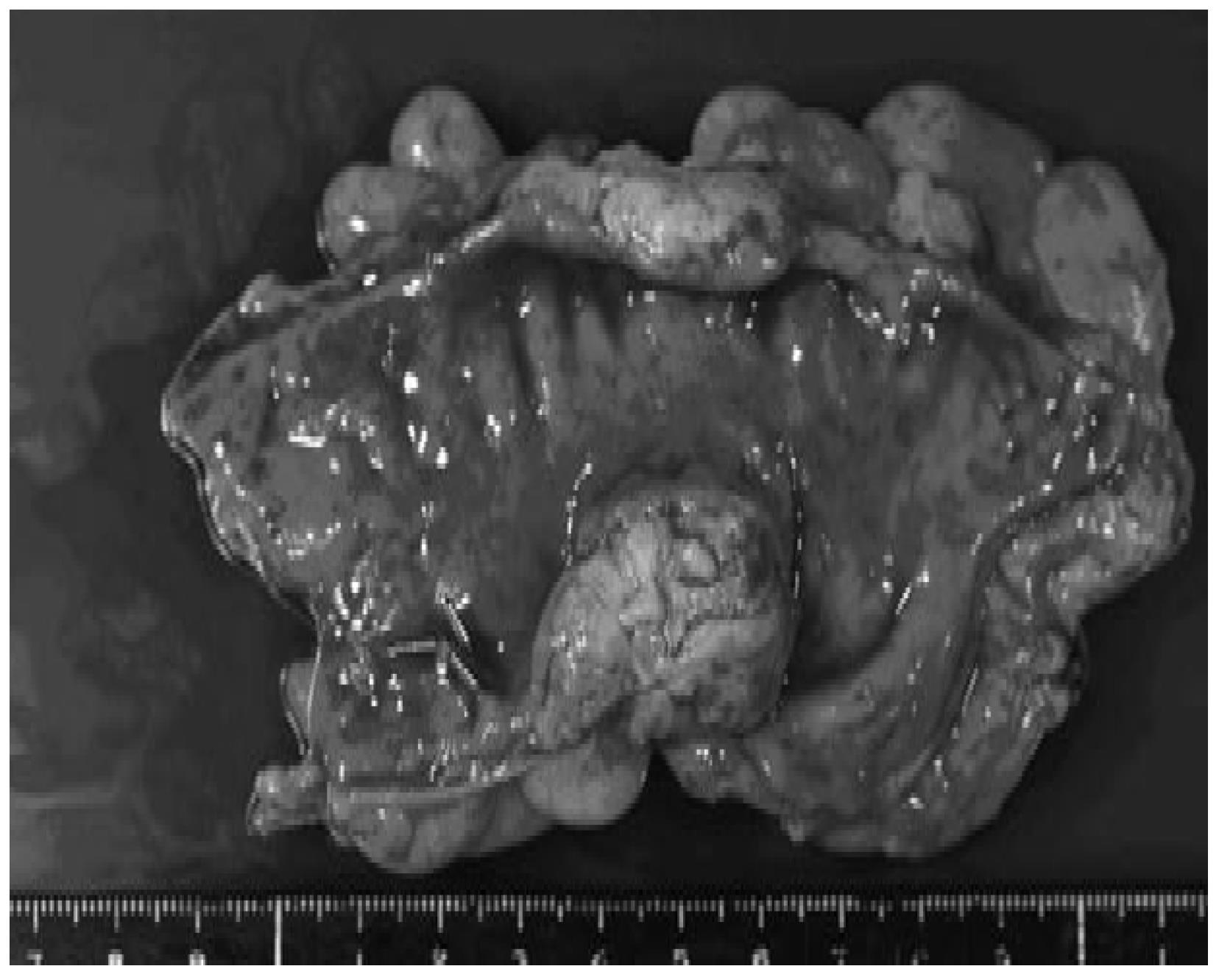
Figure 4 Gross examination of the resected specimen.
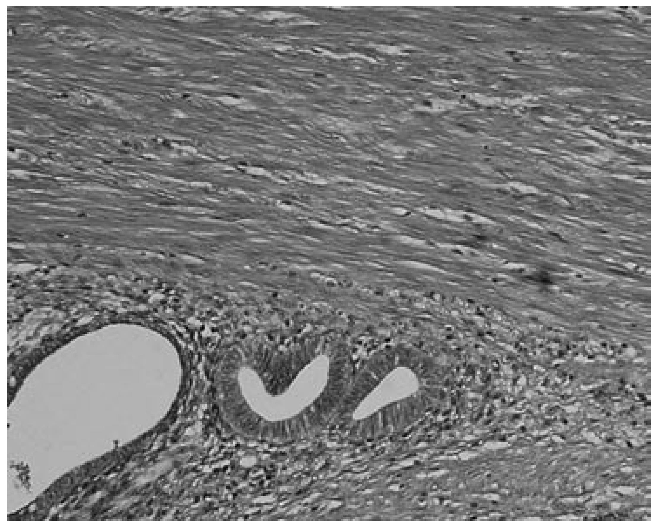
Figure 5 Tumor comprising endometrial-like grands and smooth muscle cells (× 200).
- Citation: Yoshida M, Watanabe Y, Horiuchi A, Yamamoto Y, Sugishita H, Kawachi K. Sigmoid colon endometriosis treated with laparoscopy-assisted sigmoidectomy: Significance of preoperative diagnosis. World J Gastroenterol 2007; 13(40): 5400-5402
- URL: https://www.wjgnet.com/1007-9327/full/v13/i40/5400.htm
- DOI: https://dx.doi.org/10.3748/wjg.v13.i40.5400


