Copyright
©2007 Baishideng Publishing Group Co.
World J Gastroenterol. Oct 28, 2007; 13(40): 5312-5316
Published online Oct 28, 2007. doi: 10.3748/wjg.v13.i40.5312
Published online Oct 28, 2007. doi: 10.3748/wjg.v13.i40.5312
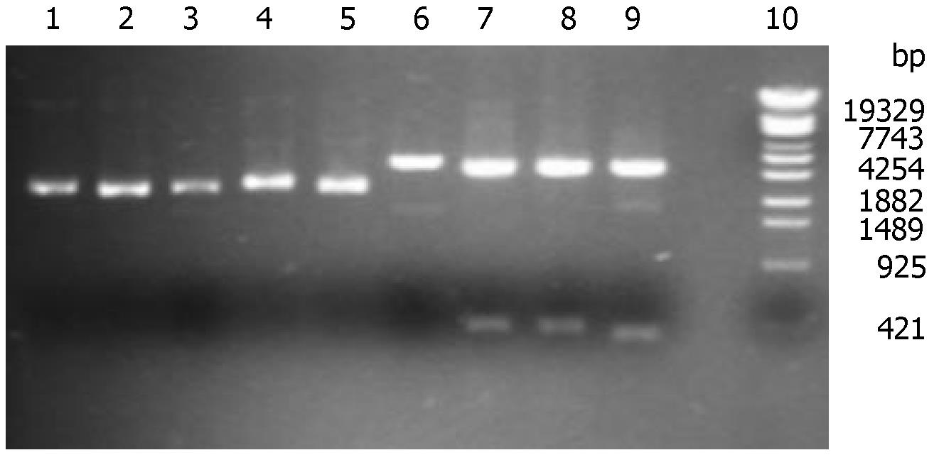
Figure 1 Restriction map of recombinant plasmid pShRNA.
Lanes 1-2: pShRNA undigested; Lane 3: undigested null vector pTZU6+1; Lanes 4-5: pShRNA digested by salI; Lane 6: pTZU6+1 digested by salI; Lanes 7-8: pShRNA digested by Hind III and EcoRI(2800 + 395 bp); Lane 9: pTZU6+1 digested by Hind III and EcoRI(2800 + 352 bp); Lane 10: λT14 DNA Marker.
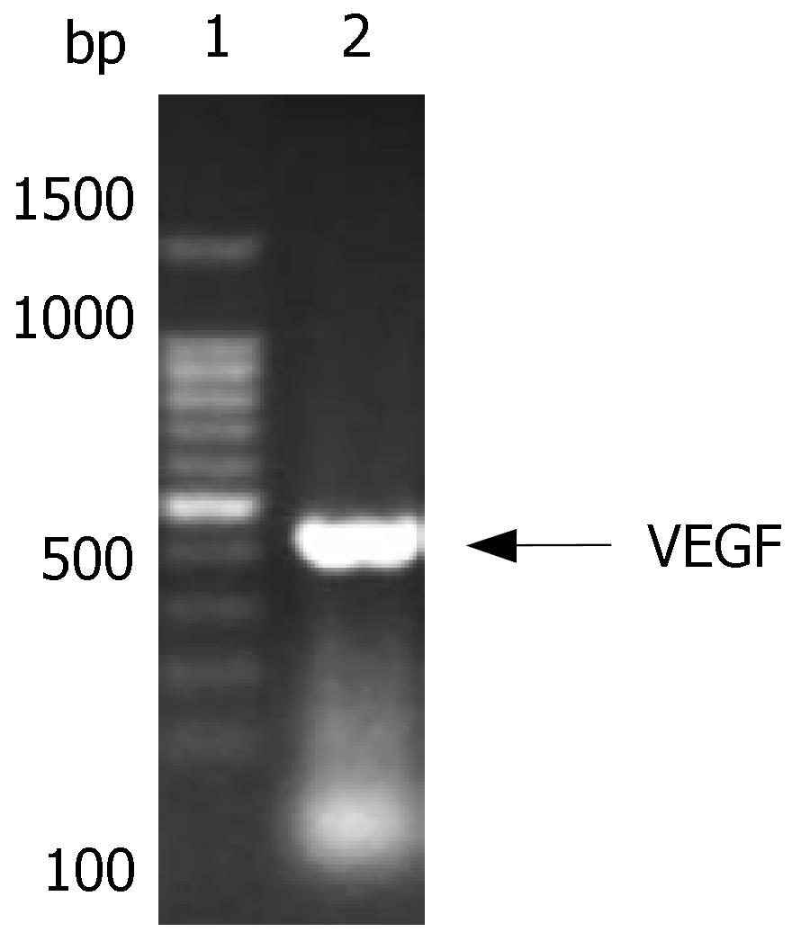
Figure 2 Gel electrophoresis of VEGF.
Lane 1: 100 bp Marker; Lane 2: RT-PCR for VEGF (411 bp).
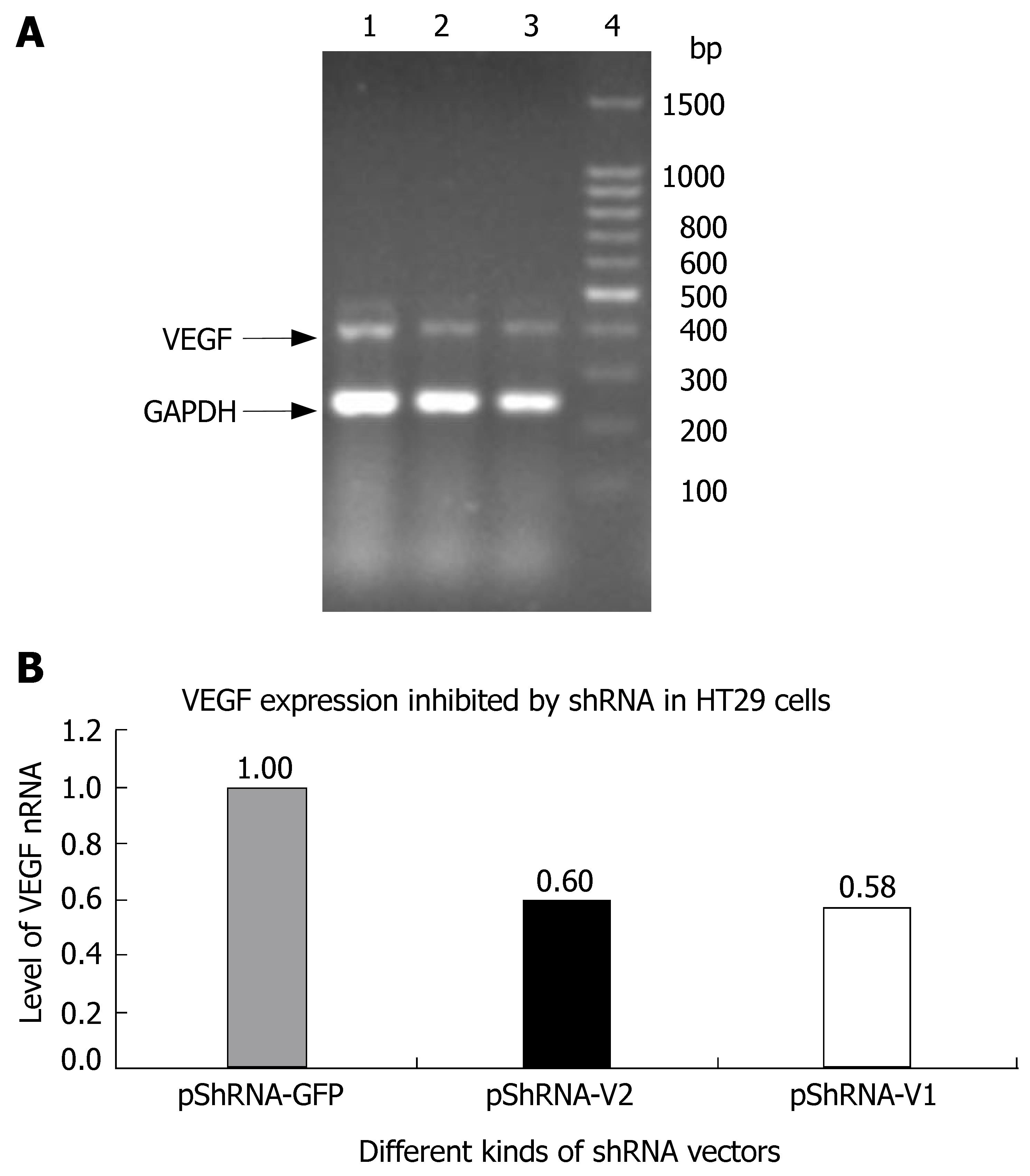
Figure 3 VEGF gene expression inhibited by shRNAs (A) and the inhibition rate of pShRNA-V1 and pShRNA-V2 (B) in HT29 cells.
Lane 1: pShRNA-GFP; Lane 2: pShRNA-V2; Lane 3: pShRNA-V1; Lane 4: 100 bp Marker.
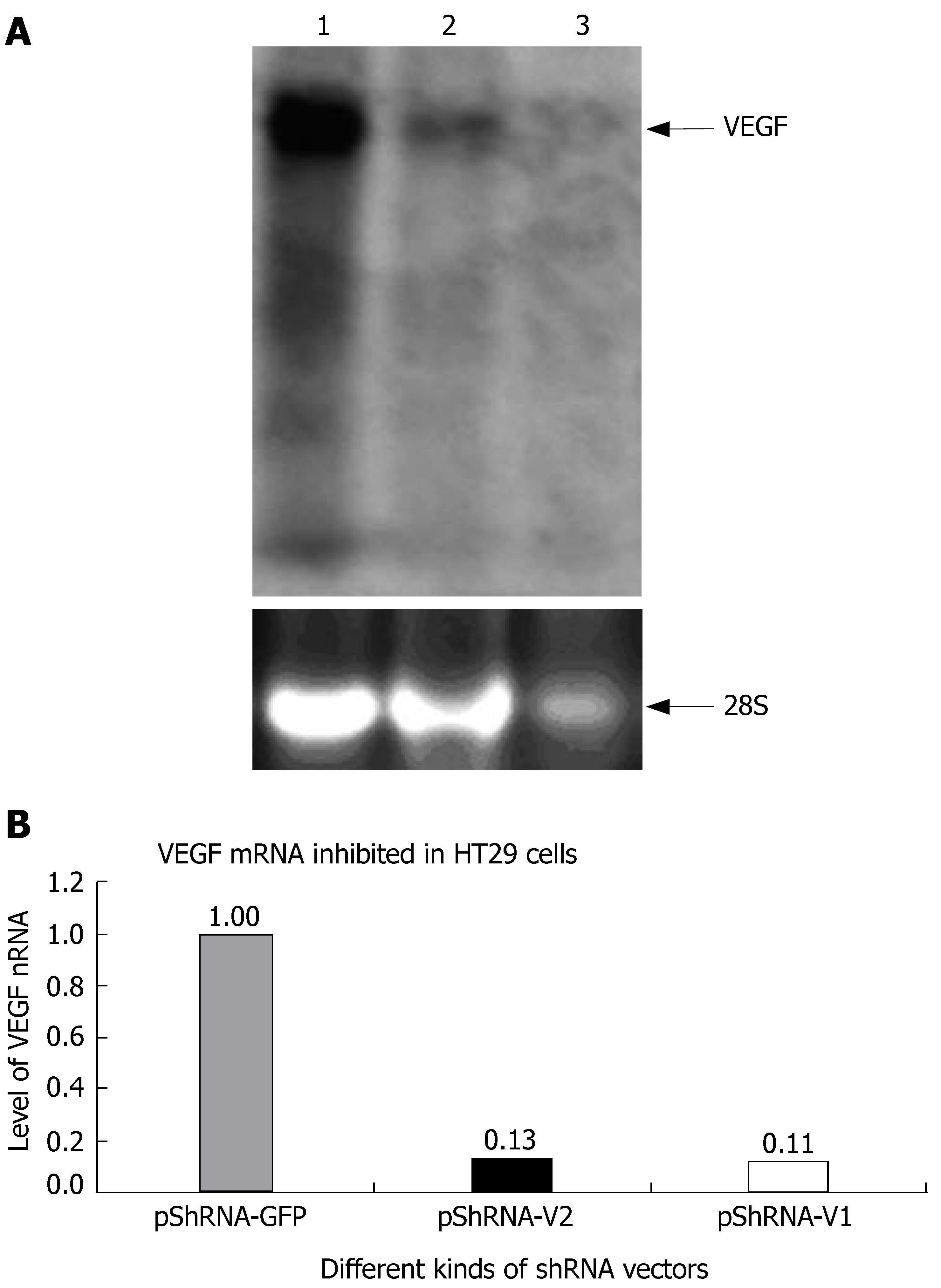
Figure 4 VEGF mRNA inhibited in HT29 cells by Northern blotting analysis (A) and the inhibition rate of pShRNA-V1 and pShRNA-V2 (B).
Lane 1: pShRNA-GFP; Lane 2: pShRNA-V1; Lane3: pShRNA-V2.
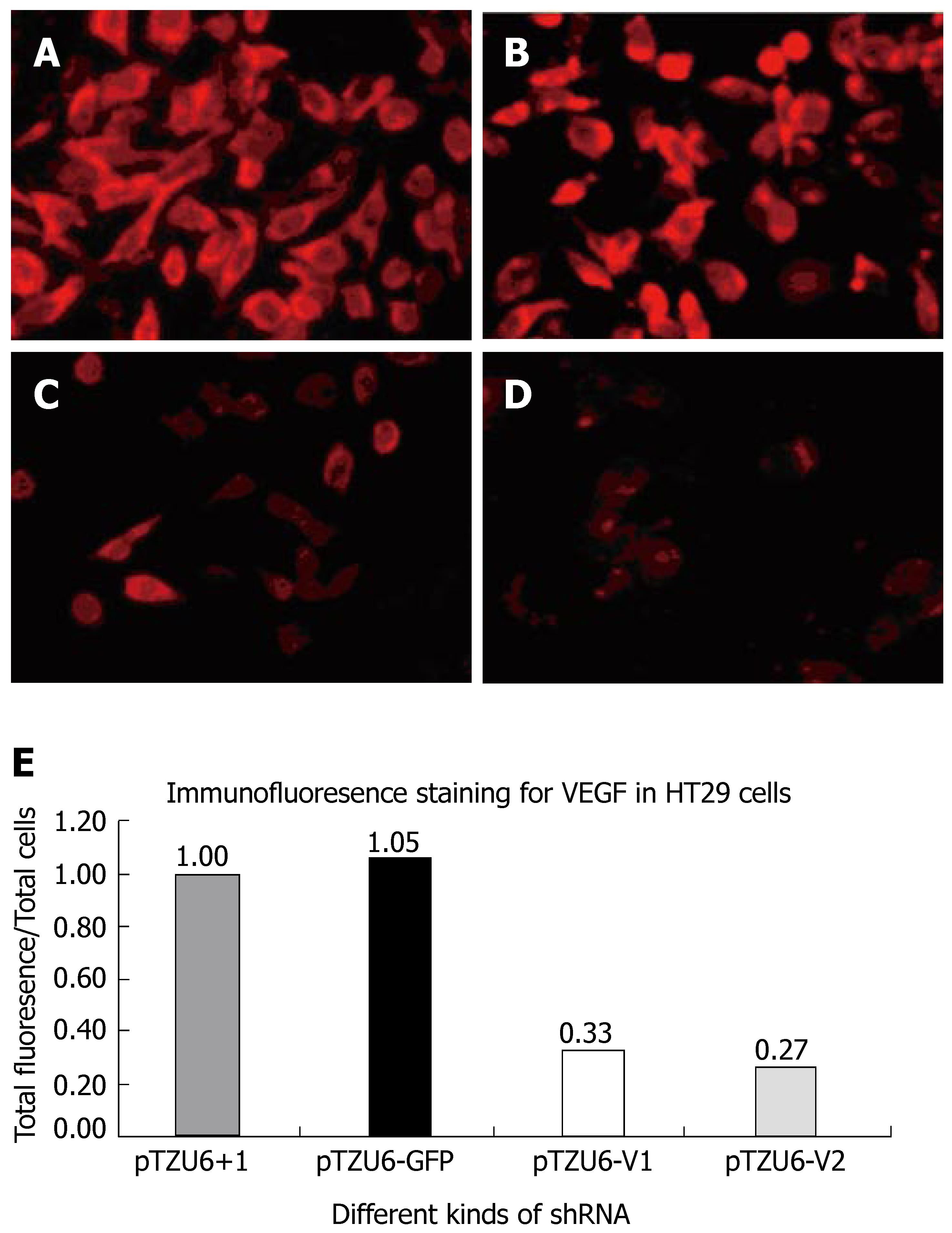
Figure 5 Immunofluoresence staining for pTZU6+1 (A), pTZU6-GFP (B), pTZU6-V1 (C), pTZU6-V2 (D), and the inhibition rate of pShRNA-V1 and pShRNA-V2 (E).
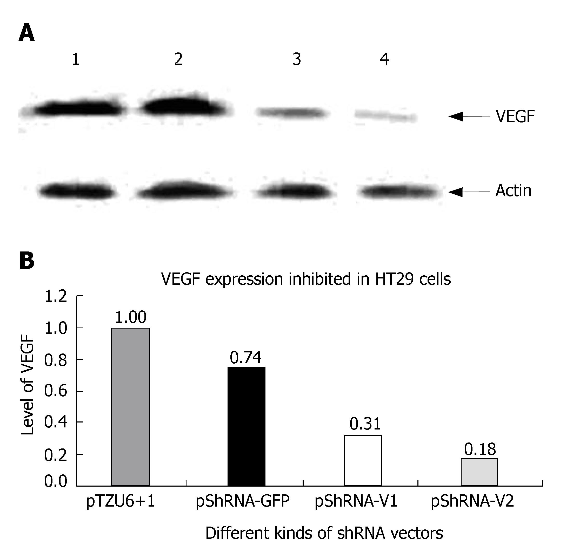
Figure 6 Western blotting for VEGF (A) and the inhibition rate of pShRNA-V1 and pShRNA-V2 (B) in HT29 cells.
Lane 1: pTZU6+1; Lane 2: pShRNA-G; Lane 3: pShRNA-V1; Lane 4: pShRNA-V2.
- Citation: Li TJ, Song JN, Kang K, Tong SS, Hu ZL, He TC, Zhang BQ, Zhang CQ. RNA interference-mediated gene silencing of vascular endothelial growth factor in colon cancer cells. World J Gastroenterol 2007; 13(40): 5312-5316
- URL: https://www.wjgnet.com/1007-9327/full/v13/i40/5312.htm
- DOI: https://dx.doi.org/10.3748/wjg.v13.i40.5312


