Copyright
©2007 Baishideng Publishing Group Inc.
World J Gastroenterol. Oct 21, 2007; 13(39): 5232-5237
Published online Oct 21, 2007. doi: 10.3748/wjg.v13.i39.5232
Published online Oct 21, 2007. doi: 10.3748/wjg.v13.i39.5232
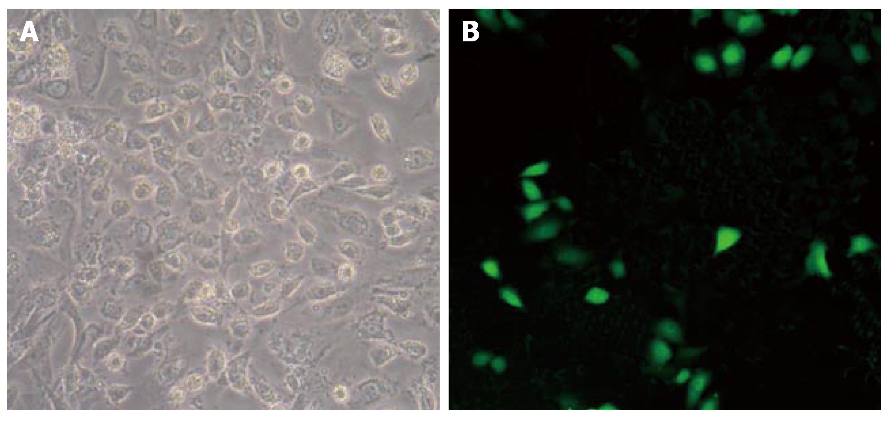
Figure 1 Transfection of recombination plasmids into cultured (A) and non-cultured (B) pancreatic ductal epithelial cells.
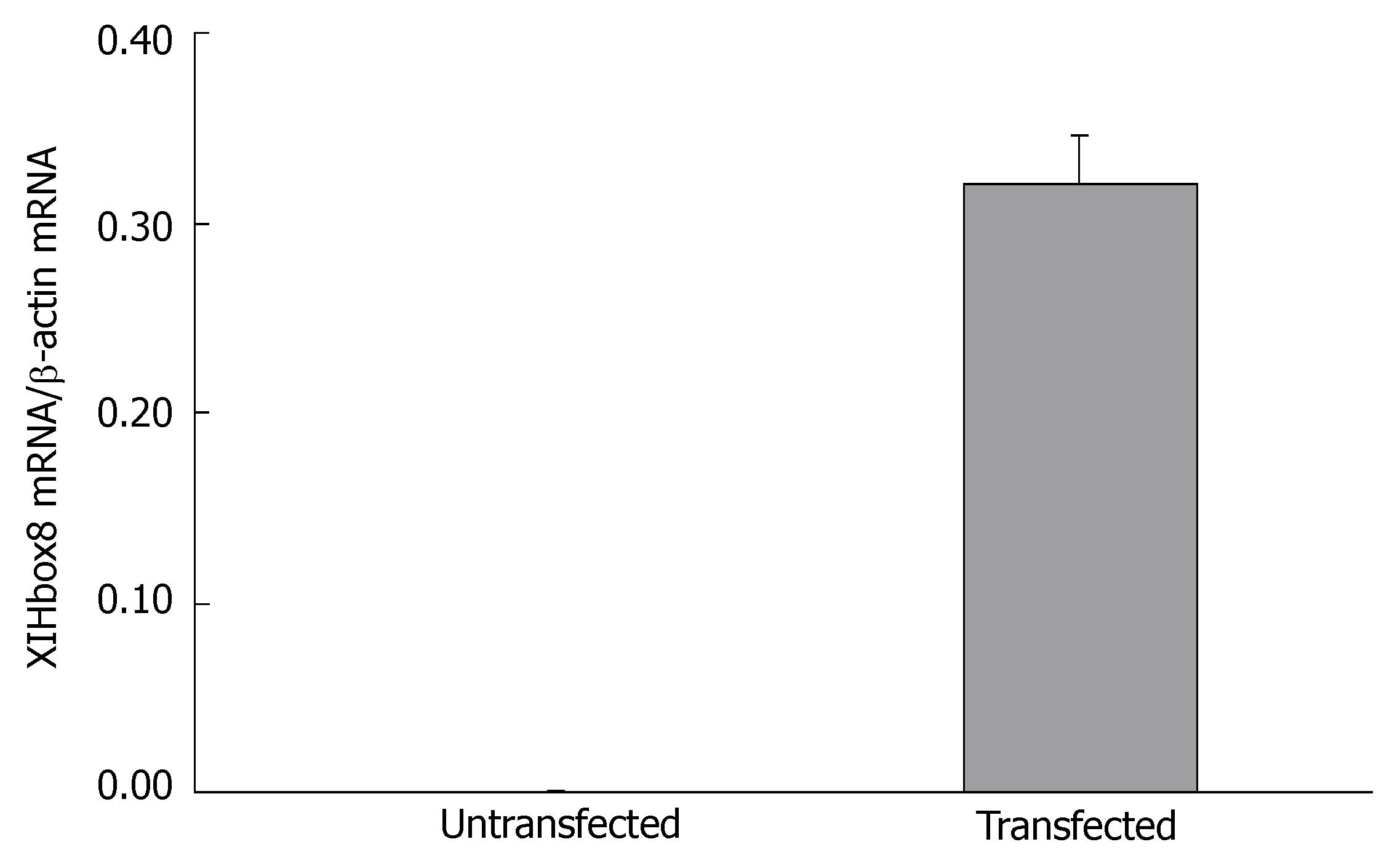
Figure 2 XlHbox8 mRNA expression 24 h after transfection of recombination plasmids.
Figure 3 PDX-1 and insulin mRNA expression after transfection of recombination plasmids.
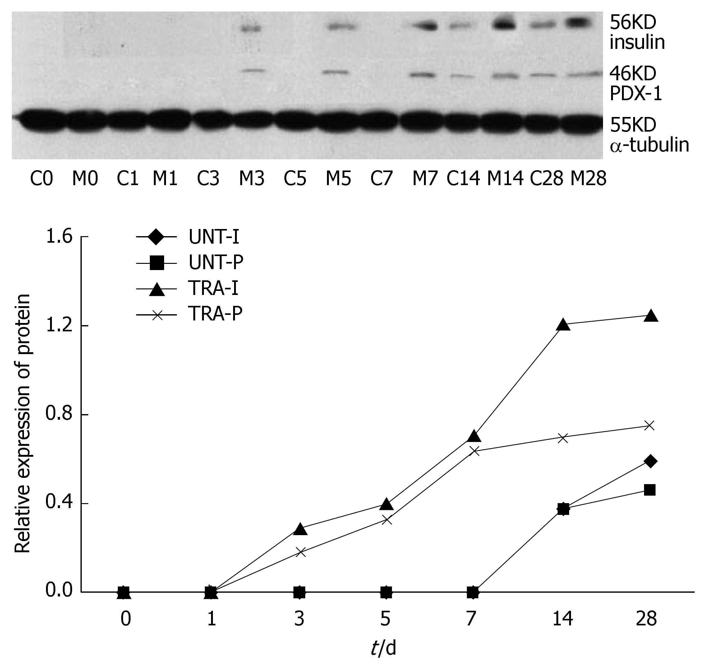
Figure 4 Western blot analysis of PDX-1 and insulin protein showing the increased expression of PDX-1 (P < 0.
01) and insulin (P < 0.01) protein in the transfected cells. C0-C28: Untransfected group on d 0-28; M0-M28: Transfected group on d 0-28; UNT-I: Insulin expression in untransfected group; UNT-P: PDX-1 expression in untransfected group; TRA-I: Insulin expression in transfected group; TRA-P: PDX-1 expression in transfected group.
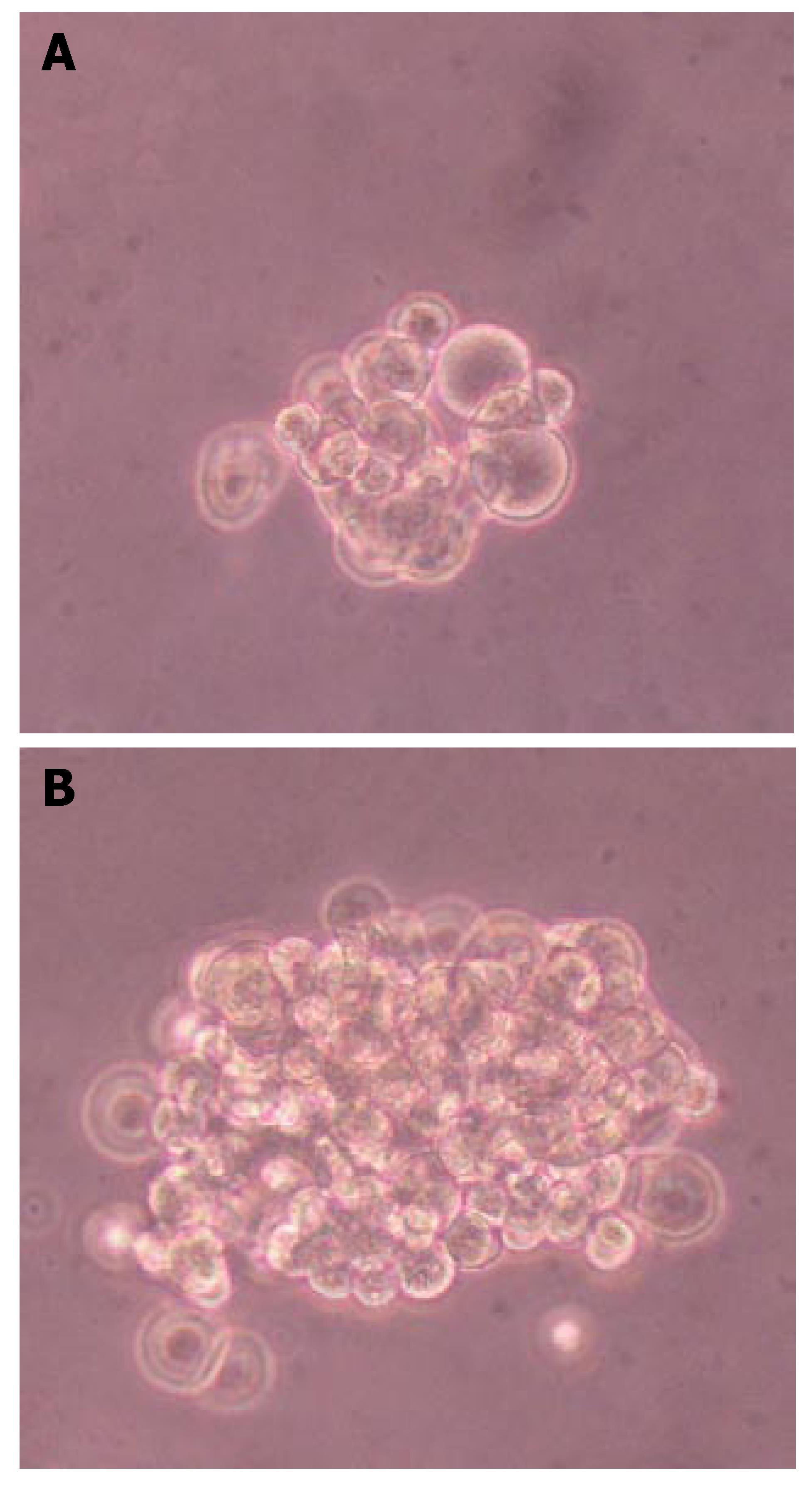
Figure 5 Dithizone staining of insulin-producing cells in untransfected group (A) and transfected group (B).
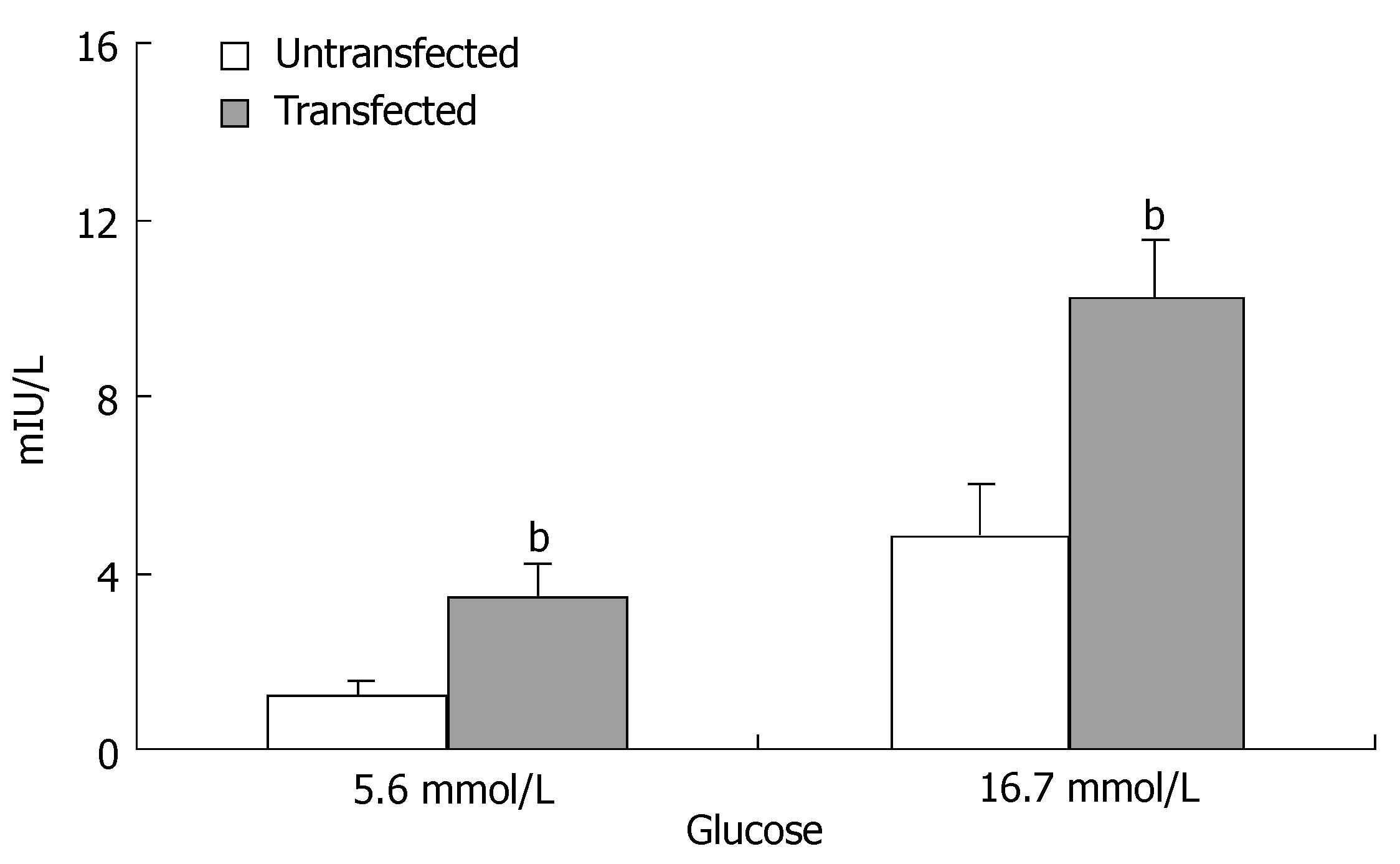
Figure 6 Response of insulin secretion to physiological stimuli.
Insulin secretion was significantly increased in the transfected group compared with the untransfected group (bP < 0.01).
-
Citation: Liu T, Wang CY, Yu F, Gou SM, Wu HS, Xiong JX, Zhou F.
In vitro pancreas duodenal homeobox-1 enhances the differentiation of pancreatic ductal epithelial cells into insulin-producing cells. World J Gastroenterol 2007; 13(39): 5232-5237 - URL: https://www.wjgnet.com/1007-9327/full/v13/i39/5232.htm
- DOI: https://dx.doi.org/10.3748/wjg.v13.i39.5232


