Copyright
©2007 Baishideng Publishing Group Co.
World J Gastroenterol. Jul 21, 2007; 13(27): 3767-3769
Published online Jul 21, 2007. doi: 10.3748/wjg.v13.i27.3767
Published online Jul 21, 2007. doi: 10.3748/wjg.v13.i27.3767
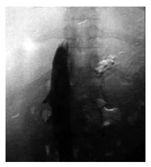
Figure 1 Venography of complete obstruction of IVC.
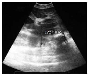
Figure 2 Location of venous thrombosis in IVC on the ultrasound.
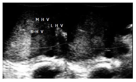
Figure 3 Post-stenting sonography, showing the right, left and middle hepatic veins.
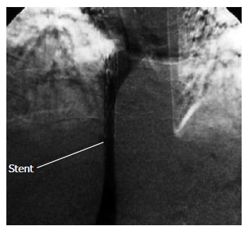
Figure 4 Post-stenting venography.
- Citation: Reza F, Naser DE, Hossein G, Mehrdad Z. Combination of thrombolytic therapy and angioplastic stent insertion in a patient with Budd-Chiari syndrome. World J Gastroenterol 2007; 13(27): 3767-3769
- URL: https://www.wjgnet.com/1007-9327/full/v13/i27/3767.htm
- DOI: https://dx.doi.org/10.3748/wjg.v13.i27.3767


