Copyright
©2007 Baishideng Publishing Group Co.
World J Gastroenterol. May 7, 2007; 13(17): 2490-2495
Published online May 7, 2007. doi: 10.3748/wjg.v13.i17.2490
Published online May 7, 2007. doi: 10.3748/wjg.v13.i17.2490
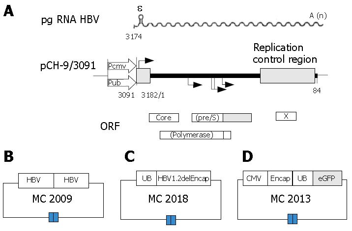
Figure 1 Schematic representation of plasmid constructs.
A: Plasmid constructs used for the production of recombinant hepatitis B virus (rHBV)[10], (pgRNA HBV = sinusoidal line; ε = encapsidation signal; A (n) = poly A tail; CMV = cytomegalovirus; UB= ubiquitin); B: The vector MC2009 contains 2 tandem copies of HBV genome; C: The transfer plasmid DNA vector MC2013 obtained by replacing a DNA fragment containing the small envelope gene (HBV position 1446 to 2347) with a PCR fragment encoding a fluorescence-enhanced green fluorescence protein (eGFP), meanwhile, a DNA fragment encompassing the HBV preS2/S promoter was substituted by a PCR fragment encoding UB promoter which could drive transgene expression. A CMV promoter was introduced to drive the expression of the UB.eGFP reporter expression cassette with the HBV encapsidation signal, and premature stop codons were introduced into all remaining HBV open reading frames; D: The helper plasmid MC2018 contains only one copy of the HBV genome with a deletion of the encapsidation signal ε or direct repeats necessary for reverse transcription. The UB promoter was introduced to drive expression of HBV 1.2delEncap.
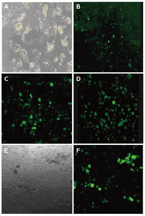
Figure 2 Transduction of primary human hepatocytes (PHH) by the HBV-based vector.
PHH were infected with rHBV-GFP, a recombinant HBV that carries a GFP gene under control of the UB promoter (described previously in Figure 1). A: Cell morphology of PHH on d 30; B: GFP expression on d 3 post-infection; C: GFP expression on d 6 post-infection; D: on d 12 post-infection; E: Transduction of PHH demonstrated by an overlay of the fluorescence; F: Phase contrast of the same field.
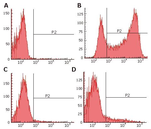
Figure 3 PHH were detected by FACS.
The purity of PHH cultures was analyzed by identification of nonparenchymal and parenchymal liver cells on d 6 after seeding. A: Control; B: FACS analysis of parenchymal liver cells with GFP; C: FACS analysis of kupffer cells with Dextran conjugates; D: FACS analysis of liver sinusoidal endothelial cells (LSEC) with acetylated low-density lipoprotein. The population consisted of 10.4% AcLDL positive cells, 2% Carboxy-Q-Rhodamine positive cells and 75.2% GFP positive cells, meaning that total parenchymal cells is about 87.6%, approximately 85% of it were infected by HBV based vector with GFP.
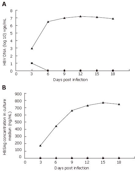
Figure 4 The efficiency of PHH infected with wt HBV or rHBV.
PHH were infected at a multiplicity of infection of 10 DNA-containing HBV particles/cell on d 1 post-seeding, HBV DNA (A), and HBSAg (B) secreted into cell culture medium were determined every 3 d after infection. The triangles denote PHH cells infected with wt HBV, and squares denote PHH cells infected with rHBV. In the supernatant from PHH infected with rHBV, no HBV DNA and HBsAg can be detected, meaning that all viral gene were knocked out.
- Citation: Li SH, Huang WG, Huang B, Chen XG. High expression of hepatitis B virus based vector with reporter gene in hepatitis B virus infection system. World J Gastroenterol 2007; 13(17): 2490-2495
- URL: https://www.wjgnet.com/1007-9327/full/v13/i17/2490.htm
- DOI: https://dx.doi.org/10.3748/wjg.v13.i17.2490


