Copyright
©2007 Baishideng Publishing Group Co.
World J Gastroenterol. May 7, 2007; 13(17): 2490-2495
Published online May 7, 2007. doi: 10.3748/wjg.v13.i17.2490
Published online May 7, 2007. doi: 10.3748/wjg.v13.i17.2490
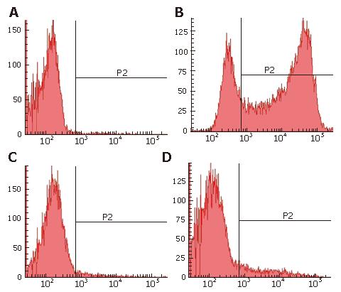
Figure 3 PHH were detected by FACS.
The purity of PHH cultures was analyzed by identification of nonparenchymal and parenchymal liver cells on d 6 after seeding. A: Control; B: FACS analysis of parenchymal liver cells with GFP; C: FACS analysis of kupffer cells with Dextran conjugates; D: FACS analysis of liver sinusoidal endothelial cells (LSEC) with acetylated low-density lipoprotein. The population consisted of 10.4% AcLDL positive cells, 2% Carboxy-Q-Rhodamine positive cells and 75.2% GFP positive cells, meaning that total parenchymal cells is about 87.6%, approximately 85% of it were infected by HBV based vector with GFP.
- Citation: Li SH, Huang WG, Huang B, Chen XG. High expression of hepatitis B virus based vector with reporter gene in hepatitis B virus infection system. World J Gastroenterol 2007; 13(17): 2490-2495
- URL: https://www.wjgnet.com/1007-9327/full/v13/i17/2490.htm
- DOI: https://dx.doi.org/10.3748/wjg.v13.i17.2490


