Copyright
©2007 Baishideng Publishing Group Co.
World J Gastroenterol. Apr 7, 2007; 13(13): 2004-2006
Published online Apr 7, 2007. doi: 10.3748/wjg.v13.i13.2004
Published online Apr 7, 2007. doi: 10.3748/wjg.v13.i13.2004
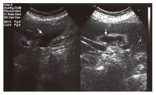
Figure 1 Oblique and sagittal US images showing two lumens.
The first (1) has a normal wall thickness and lumen; the second (2) has a thickened wall (arrow) and is filled with sludge (arrowheads).
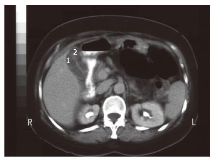
Figure 2 Transverse CT image with 10-mm slice thickness and interval showing two lumens with normal (1), and thickened (2) walls.
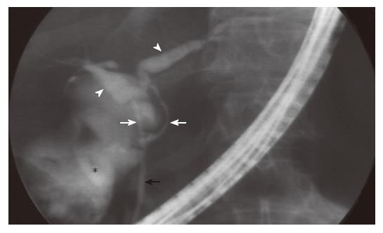
Figure 3 ERCP shows two hepatic ducts (arrowheads), two cystic ducts (white arrows) and the common bile duct (black arrow).
None of the gallbladder lumens were opacified since accompanying inflammation obstruct the cystic duct. The opacity shown by asterix is the contrast material leak into the duodenum lumen.
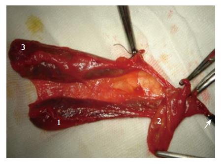
Figure 4 Pathologic specimen shows three distinct gallbladders each bearing a separate cystic duct and all joining to the common bile duct (arrow).
Bladder number 1 and 3 were normal, bladder number 3 was inflammated.
- Citation: Alicioglu B. An incidental case of triple gallbladder. World J Gastroenterol 2007; 13(13): 2004-2006
- URL: https://www.wjgnet.com/1007-9327/full/v13/i13/2004.htm
- DOI: https://dx.doi.org/10.3748/wjg.v13.i13.2004


