Copyright
©2007 Baishideng Publishing Group Co.
World J Gastroenterol. Apr 7, 2007; 13(13): 1975-1979
Published online Apr 7, 2007. doi: 10.3748/wjg.v13.i13.1975
Published online Apr 7, 2007. doi: 10.3748/wjg.v13.i13.1975
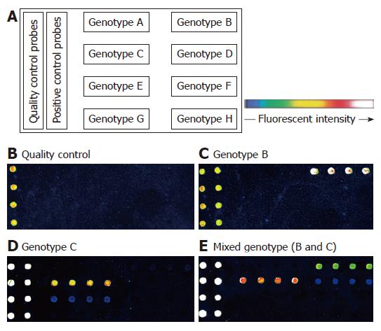
Figure 1 Layout of probes and fluorescence images of results.
Probes layout on the oligonucleotide chip (A). Following hybridization of Cy5-labeled fragmented DNA, as described in Materials and Methods, the fluorescent signals of different HBV genotypes were detected as follows: Genotype B (C), genotype C (D), mixed genotype (E). Blank template amplified by PCR also was hybridized to the chip and the image of the result is shown in (B).
- Citation: Tang XR, Zhang JS, Zhao H, Gong YH, Wang YZ, Zhao JL. Detection of hepatitis B virus genotypes using oligonucleotide chip among hepatitis B virus carriers in Eastern China. World J Gastroenterol 2007; 13(13): 1975-1979
- URL: https://www.wjgnet.com/1007-9327/full/v13/i13/1975.htm
- DOI: https://dx.doi.org/10.3748/wjg.v13.i13.1975


