Copyright
©2007 Baishideng Publishing Group Co.
World J Gastroenterol. Apr 7, 2007; 13(13): 1947-1952
Published online Apr 7, 2007. doi: 10.3748/wjg.v13.i13.1947
Published online Apr 7, 2007. doi: 10.3748/wjg.v13.i13.1947
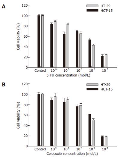
Figure 1 Viability of colon cancer cells treated with 5-FU and celecoxib.
Cell viability was determined by MTT assay. Cell viabilities at various concentrations of 5-FU (A) and celecoxib (B) were evaluated using a cell viability index (%), which was defined as: (mean absorbance in the test group/mean absorbance in the control group) x 100. Black bars represent HCT-15 cells and white bars HT-29 cells. Results are mean ± SE.
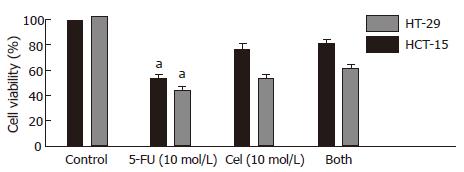
Figure 2 Combinatorial chemotherapy of HCT-15 or HT-29 human colon cancer cells with 5-FU and/or celecoxib.
Cells were treated with 10-3 mol/L 5-FU, 10-5 mol/L celecoxib, or both. Cell viability was determined by the MTT assay, and expressed as the cell viability index (%) defined as: (mean absorbance in the test group/mean absorbance in the control group) × 100. Results are mean ± SE. aP < 0.05 vs Both.
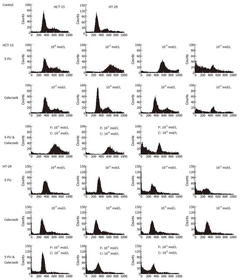
Figure 3 Flow cytometry of human colon cancer cells.
Cells were treated with various concentrations of 5-FU, celecoxib, and both drugs. The cells were washed once with PBS, centrifuged and stained with propidium iodide, and then analyzed using a FACSCalibur. F represents 5-FU, and C celecoxib.
Figure 4 Western blotting of apoptosis molecules.
A: Control; B: 10-3 mol/L 5-FU; C: 10-5 mol/L Celecoxib; D: 5-FU (10-3 mol/L) and celecoxib (10-5 mol/L). HCT-15 and HT-29 human colon cancer cell lines were treated with 10-3 mol/L 5-FU, 10-5 mol/L celecoxib, and 5-FU (10-3 mol/L) and celecoxib (10-5 mol/L). Western blotting for apoptosis-related molecules was performed as described in ‘Materials and Methods’. Co-treatment with celecoxib attenuated the caspase-3 expression and PARP cleavage.
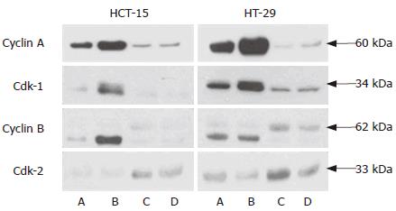
Figure 5 Western blotting of cell cycle-regulatory molecules.
A: Control; B: 10-3 mol/L 5-FU; C: 10-5 mol/L Celecoxib; D: 5-FU (10-3 mol/L ) and celecoxib (10-5 mol/L ). HCT-15 and HT-29 human colon cancer cells were treated with 10-3 mol/L 5-FU, 10-5 mol/L celecoxib, and the two in combination. Western blotting for cdk1, cyclin A, cyclin B, and cdk2 were performed as described in ‘Materials and Methods’. Celecoxib reduced G2/M phase accumulation, and increased the G1/S phase protein, cdk-2.
- Citation: Lim YJ, Rhee JC, Bae YM, Chun WJ. Celecoxib attenuates 5-fluorouracil-induced apoptosis in HCT-15 and HT-29 human colon cancer cells. World J Gastroenterol 2007; 13(13): 1947-1952
- URL: https://www.wjgnet.com/1007-9327/full/v13/i13/1947.htm
- DOI: https://dx.doi.org/10.3748/wjg.v13.i13.1947


