Copyright
©2007 Baishideng Publishing Group Co.
World J Gastroenterol. Apr 7, 2007; 13(13): 1906-1911
Published online Apr 7, 2007. doi: 10.3748/wjg.v13.i13.1906
Published online Apr 7, 2007. doi: 10.3748/wjg.v13.i13.1906
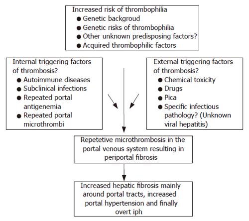
Figure 1 Our proposed theory about etiopathogenesis of idiopathic portal hypertension.
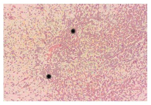
Figure 2 The histological examination of the portal vein shows periportal thickening (marked by asterisks) found in an IPH patient.
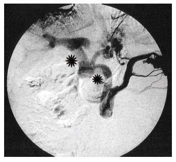
Figure 3 Splenopo-rtography shows two major sites of thrombosis in portal venous system (shown by asterisks).
Note the dilated portal vein at hepatic hilum despite portal vein thrombosis.
Figure 4 The computed tomography of the same patient in figure-3.
Note the thrombosis in the portal vein (Marked by arrow).
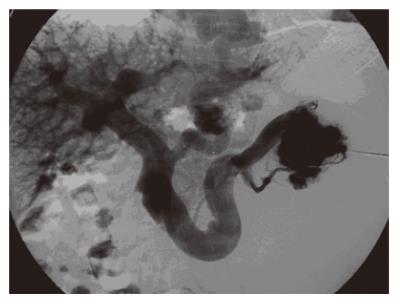
Figure 5 Splenoportography shows patent portal vein with severe dilation and collateral circulation.
Note the small caliber of intrahepatic portal vein branches compared to extrahepatic portal vein.
- Citation: Harmanci O, Bayraktar Y. Clinical characteristics of idiopathic portal hypertension. World J Gastroenterol 2007; 13(13): 1906-1911
- URL: https://www.wjgnet.com/1007-9327/full/v13/i13/1906.htm
- DOI: https://dx.doi.org/10.3748/wjg.v13.i13.1906


