Copyright
©2007 Baishideng Publishing Group Co.
World J Gastroenterol. Mar 14, 2007; 13(10): 1541-1546
Published online Mar 14, 2007. doi: 10.3748/wjg.v13.i10.1541
Published online Mar 14, 2007. doi: 10.3748/wjg.v13.i10.1541
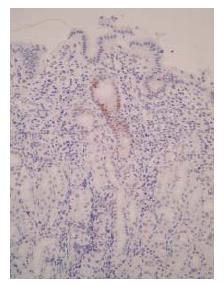
Figure 1 Immunohistochemistry for p53 (DO-7) in H pylori-infected gastric mucosa.
Positive staining for p53 (DO-7) was observed in the gastric pit, especially in the neck region before eradication (× 200).
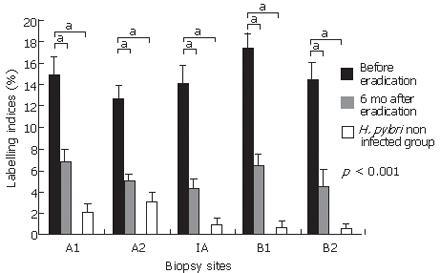
Figure 2 Labeling indices for p53 (DO-7) in 42 patients with H pylori infection, 6 mo after eradication, and 11 patients without H pylori infection.
Results were shown as mean ± SEM. p53 (DO-7) indices were significantly reduced 6 mo after eradication at all biopsy sites. p53 indices for the H pylori-infected mucosa were significantly higher than those for the gastric mucosa without H pylori infection at all biopsy sites. bP < 0.001, vs H pylori non infected group.
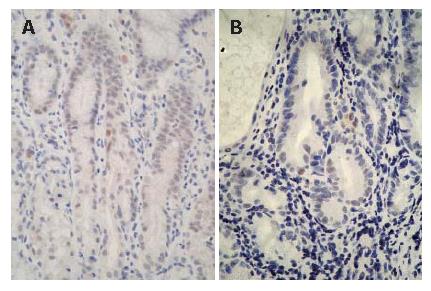
Figure 3 Immunohistochemistry for p53 (PAb240) in the H pylori-infected gastric mucosa.
A small number of cells clearly showing the expression of p53 (PAb240) were detected in the neck region before eradication. A: a 53-yr-old male with gastric ulcer; B: a 51-yr-old male with chronic gastritis; × 400).
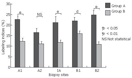
Figure 4 Labeling indices for p53 (DO-7) in group A with more than 6 positive cells for p53 (PAb240) per 10 gastric pits (n = 12) and group B with less than 5 positive cells for p53 (PAb240) per 10 gastric pits (n = 30) before eradication.
Results are shown as mean ± SEM. Labeling indices for p53 (DO-7) were significantly higher for group A than for group B at all biopsy sites except for the lesser curvature of the antrum. aP < 0.05, bP < 0.01, NS, not statistical.
-
Citation: Kodama M, Murakami K, Okimoto T, Sato R, Watanabe K, Fujioka T. Expression of mutant type-
p 53 products inH pylori -associated chronic gastritis. World J Gastroenterol 2007; 13(10): 1541-1546 - URL: https://www.wjgnet.com/1007-9327/full/v13/i10/1541.htm
- DOI: https://dx.doi.org/10.3748/wjg.v13.i10.1541


